. 2010 Jan;40(1):134-41.
doi: 10.1002/eji.200939258.

A novel human CD4+ T-cell inducer subset with potent immunostimulatory properties

Affiliations

A novel human CD4+ T-cell inducer subset with potent immunostimulatory properties

Lishomwa C Ndhlovu et al. Eur J Immunol. 2010 Jan.

Abstract

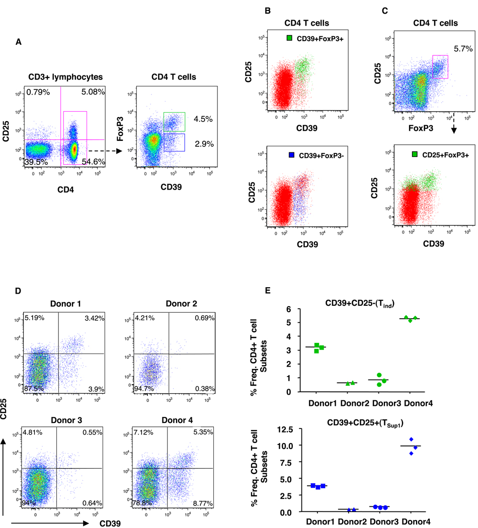
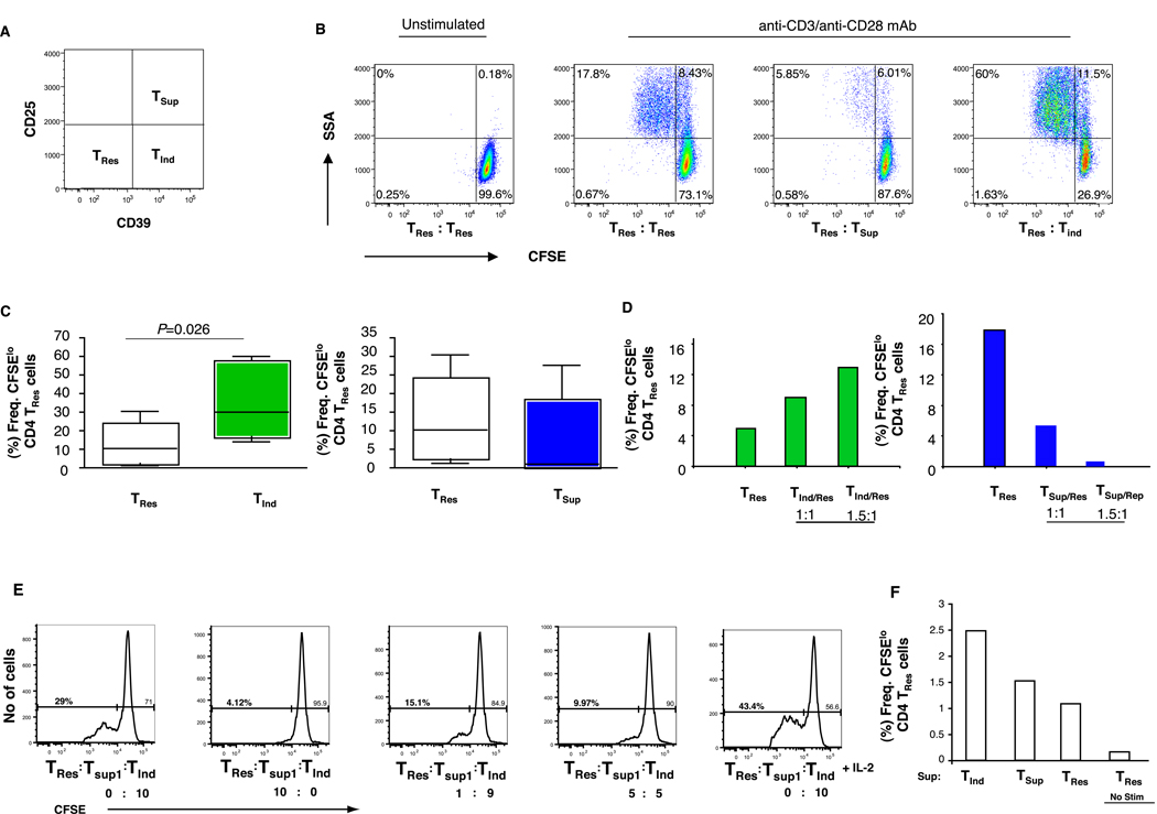
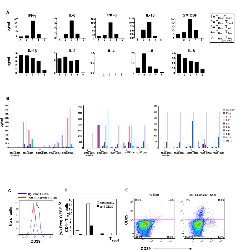
The complexity of immunoregulation has focused attention on the CD4+ T "suppressor" regulatory cell (Treg), which helps maintain balance between immunity and tolerance. An immunoregulatory T-cell population that upon activation amplifies cellular immune responses was described in murine models more than 30 years ago; however, no study has yet identified a naturally occurring T "inducer" cell type. Here, we report that the ectoenzyme CD39/NTPDase1 (ecto-nucleoside triphosphate diphosphohydrolase 1) helps to delineate a novel population of human "inducer" CD4+ T cells (Tind) that significantly increases the proliferation and cytokine production of responder T cells in a dose-dependent manner. Furthermore, this unique Tind subset produces a distinct repertoire of cytokines in comparison to the other CD4+ T-cell subsets. We propose that this novel CD4+ T-cell population counterbalances the suppressive activity of suppressor Treg in peripheral blood and serves as a calibrator of immunoregulation.


Conflict of interest statement

There are no financial/commercial conflicts of interests.

Figures

Figure 1
Figure 1
Differential expression of CD39 on CD4+ T cell subsets. PMBC from healthy donors were surface stained for CD4, CD3, CD25, CD127 and CD39 followed by intracellular staining for FoxP3. (A) Representative dot plots showing expression of CD39 as assessed by flow cytometry on live CD3 gated lymphocytes. The frequency (%) of CD4 T cells gated is shown according to their dual expression of CD39 and FOXP3. (B) Two distinct CD39-expressing CD4 T cell subsets are identified and further delineated by CD25 and CD39 expression: CD39+FOXP3+CD25hi/+(green) and CD39+FoxP3-CD25- T cells (blue). (C) FoxP3+CD25+ gated CD4 T cells (green) were further characterized by CD25 and CD39 expression. (D) Plot shows the frequency of CD4+ T cell subsets defined by CD39 and CD25 in four representative healthy donors. (E) Stability of CD39+CD25- and CD39+CD25+ T cells as a percentage of total CD4 T cells among 4 representative healthy donors at 2–3 time points approximately 4–6 months apart. At least 200,000 lymphocyte events were counted and collected for each sample.
Figure 2
Figure 2
Differential function of CD39 expressing CD4+ T cells. Purified CD4+ T cell subsets were isolated from PBMC from healthy donors by cell sorting to a purity >95% as demonstrated in Fig 1A. (A, B) Representative plots shows 50,000 CFSE-labeled CD39-CD25- responder (TRes) T cells co-cultured in duplicate wells together with an equal number of CD39+CD25- (TInd), CD39+CD25+ (TSup) or unlabeled control CD39-CD25- (TRes) T cells for 4–5 days in the presence of soluble anti-CD3/anti-CD28. Plots show proliferation of responder TRes cells as determined by CFSE dilution. (C) Similar results were obtained in six separate experiments from six different donors. The statistical difference was deemed significant using a Mann Whitney U test analysis if p<0.05. (D, E) In some experiments TRes cells were co-cultured at different ratios with TInd cells or TSup cells or a combination of both in the absence or presence of IL-2. Data in D, E are of a representative donor from two separate experiments with two different donors. (F) Graph shows the co-culture of Tres cells with supernatants derived from 4 day stimulated (anti-CD3/anti-CD28 mAb mixture) individual CD4+ T cells subsets from a representation donor. Similar results were observed in three separate experiments from three different donors.
Figure 3
Figure 3
Detection of human cytokines in cell supernatants in anti-CD3/anti-CD28 stimulated (A) co-cultures of responder TRes cells with either 1= CD39+CD25+ (TSup1); 2= CD39-CD25+ (TSup2), 3= CD39+CD25- (TInd); 4= CD39-CD25- (TRes); or 5= unstimulated CD39-CD25- (TRes) T cells. Data is presented from a single donor with background subtracted and is a representation of three separate experiments (from three separate donors). Standard curves were performed for each biomarker using the mixed standards provided with the kit. (B) Cell sorted, purified individual CD4+ T cell subsets alone from three donors is presented. Data is the average of duplicate wells. (C) Specificity of a purified anti-human CD39 (clone BU61) mAb. PBMC from a healthy donor were incubated with either isotype control IgG antibody (blue line) or purified anti-human CD39 (clone BU61) mAb (red line) prior to staining with a conjugated anti-CD39 mAb (clone eBioA1) antibody. (D) CFSE labeled CD39-CD25- responder (TRes) T cells co-cultured in duplicate wells together with an equal number of indicated CD4 T cell subsets in the presence of either purified anti-CD39 (clone BU61) mAb or isotype IgG control. Cells were stimulated with anti-CD3/CD28 antibody mixture and cultured for 4 days before assessment of CFSE dilution. Data is a representation of two separate experiments from two different donors. (E) Plot shows expression of CD39 and CD25 by purified cell sorted CD39-CD25- responder (TRes) obtained from a representative healthy donor following anti-CD3/anti-CD28 stimulation for 12 hours. Similar results were obtained in 4 day cultures.

References

    1. von Herrath MG, Harrison LC. Antigen-induced regulatory T cells in autoimmunity. Nat Rev Immunol. 2003;3:223–232. - PubMed
    1. Yagi H, Nomura T, Nakamura K, Yamazaki S, Kitawaki T, Hori S, Maeda M, Onodera M, Uchiyama T, Fujii S, Sakaguchi S. Crucial role of FOXP3 in the development and function of human CD25+CD4+ regulatory T cells. Int Immunol. 2004;16:1643–1656. - PubMed
    1. Mills KH. Regulatory T cells: friend or foe in immunity to infection? Nat Rev Immunol. 2004;4:841–855. - PubMed
    1. Shevach EM. Regulatory T cells in autoimmmunity*. Annu Rev Immunol. 2000;18:423–449. - PubMed
    1. Wood KJ, Sakaguchi S. Regulatory T cells in transplantation tolerance. Nat Rev Immunol. 2003;3:199–210. - PubMed

Publication types

MeSH terms