. 2009 Nov;60(11):3324-35.
doi: 10.1002/art.24877.

A novel T cell cytokine, secreted osteoclastogenic factor of activated T cells, induces osteoclast formation in a RANKL-independent manner

Affiliations

A novel T cell cytokine, secreted osteoclastogenic factor of activated T cells, induces osteoclast formation in a RANKL-independent manner

Leonard Rifas et al. Arthritis Rheum. 2009 Nov.

Abstract

Objective: Chronic T cell activation is central to the etiology of rheumatoid arthritis (RA), an inflammatory autoimmune disease that leads to severe focal bone erosions and generalized systemic osteoporosis. Previous studies have shown novel cytokine-like activities in medium containing activated T cells, characterized by potent induction of the osteoblastic production of interleukin-6 (IL-6), an inflammatory cytokine and stimulator of osteoclastogenesis, as well as induction of an activity that directly stimulates osteoclast formation in a manner independent of the key osteoclastogenic cytokine RANKL. This study was undertaken to identify the factors secreted by T cells that are responsible for these activities.

Methods: Human T cells were activated using anti-human CD3 and anti-human CD28 antibodies for 72 hours in AIM V serum-free medium to obtain T cell-conditioned medium, followed by concentration and fractionation of the medium by fast-protein liquid chromatography. Biologically active fractions were resolved using sodium dodecyl sulfate-polyacrylamide gel electrophoresis. Major bands were analyzed by mass spectrometry, and a major candidate protein was identified. This novel cytokine was cloned, and its expression was analyzed using recombinant DNA technologies.

Results: A single novel cytokine that could induce both osteoblastic IL-6 production and functional osteoclast formation in the absence of osteoblasts or RANKL and that was insensitive to the effects of the RANKL inhibitor osteoprotegerin was identified in the activated T cell-conditioned medium; this cytokine was designated secreted osteoclastogenic factor of activated T cells (SOFAT). Further analysis of SOFAT revealed that it was derived from an unusual messenger RNA splice variant coded by the threonine synthase-like 2 gene homolog, which is a conserved gene remnant coding for threonine synthase, an enzyme that functions only in microorganisms and plants.

Conclusion: SOFAT may act to exacerbate inflammation and/or bone turnover under inflammatory conditions such as RA or periodontitis and in conditions of estrogen deficiency.


Figures
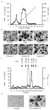
Figure 1
Figure 1
Induction of IL-6 and TRAP+ osteoclast-like cells by fractionated activated T cell conditioned medium. DEAE Sepharose column chromatography with peak fractions assayed for: (A) IL-6 (Left Y-axis) or (B) osteoclastogenic activity. Multinucleated TRAP+ cells represent osteoclasts. Control=untreated cells; Input= unfractionated T cell conditioned medium. (C) Superdex-200 fractionation of the major DEAE-sepharose peak (panel A) reveals a single peak containing both (C) IL-6 activity and (D) osteoclastogenic activity.
Figure 2
Figure 2
Nucleotide sequence, protein translation and gene structure of human SOFAT. (A) hSOFAT was cloned based on alignment of amino acid sequence derived from mass spectroscopy with hypothetical protein FLJ10916 and nucleotide sequence thus deduced. Position of 3′ and 5′ RT-PCR primers are underlined. The nucleotide sequence of hSOFAT was determined by sequencing of cDNA reverse transcribed from activated T cell total RNA and the protein sequence rederived there from. hSOFAT comprises 247 amino acids. A larger version of Part A is contained in Supplemental Figure 6. (B) Representation of how SOFAT is generated from THNSL2, using splice variant “b” (see Supplemental Figure 2) as the reference sequence. Exons shown as numbered black boxes, introns as lines (not too scale), and untranslated regions as narrow open bars. SOFAT shows highest homology to splice variant “b”, and comprises the last 97 bp of exon 4 joined to exons 5, 6, and 7, plus the first 141 bp of exon 8. An additional 33 bp of 3′UTR sequence and at least 258 bp of non coding UTR derived from an unused exon 9 is contained within the boundaries of the 3′ primer.
Figure 3
Figure 3
Expression of recombinant SOFAT, protein translation and IL-6 inducing activity. (A) E. coli expressed rhSOFAT was resolved by SDS-PAGE and Coomassie Blue stained (Lane 1), and immunoprobed with anti-polyhistidine antibody (Lane 2). (B) Protein identity was verified by MS sequencing. (C) To examine secretion, SOFAT was immunoprecipitated from CHO cell supernatant and assayed on Western blot by anti-polyhistidine antibody. Replicate assays of rhSOFAT are shown. (D) ELISA assay of rhSOFAT induced IL-6 production in osteoblasts. Data represent mean ± S.E.M. of triplicate cultures. * = p < 0.001 (One Way ANOVA,) relative to untreated cells (Untreated) and Bovine Serum Albumin (BSA) treated cell controls.
Figure 4
Figure 4
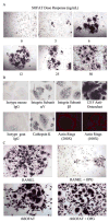
Generation and characterization of human osteoclasts by rhSOFAT. (A) Dose Response: Human monocytes were cultured with rhSOFAT for 10 days and stained for TRAP. Magnification = 100×. (B) Monocytes were cultured with rhSOFAT (100 ng/mL) for 7 days and immunostained with mouse IgG antibodies against αV and β3 integrin subunits, and the anti-osteoclast antibody 121F, or with goat IgG antibodies against cathepsin K or with the relevant mouse or goat IgG isotype controls. Magnification = 100×. Actin ring formation was visualized under fluorescence microscopy. Magnification = 100× or 200× as indicated. (C) Human monocytes were cultured with either 25 ng/mL RANKL or 100 ng/mL SOFAT ± 200 fold excess OPG for 10 days and stained for TRAP. Magnification = 100×.
Figure 5
Figure 5
rhSOFAT-induces functional osteoclasts by mouse RAW264.7 cells. (A) Mouse RAW264.7 cells were treated with rhSOFAT (100 ng/mL) for 5 days and TRAP stained. Photographed at 100× magnification. (B) To test osteoclast activity RAW264.7 cells were cultured untreated on BioCoat, or treated with rhSOFAT (100 ng/mL) or RANKL (60 ng/mL) for 10 days. Lower panel shows bright field microscopy of pits at 100× magnification. Top panel shows phase contrast microscopy of resorption pits at 200 × magnification.
Figure 6
Figure 6
SOFAT requires neither TNFα nor IL-6 for osteoclast formation however; TNFα amplifies rhSOFAT induced osteoclastogenesis. RAW264.7 cells were cultured with rhSOFAT (100 ng/mL) ± (A) TNFα (10 ng/mL); (B) neutralizing anti-TNFα antibody (20 μg/mL); or (C) anti-IL-6 antibody (20 μg/mL). After 5 days TRAP+ multinucleated cells were quantitated under light microscopy. *<0.001 vs Unstimulated; **<0.001 vs SOFAT (1-way ANOVA (Tukey-Kramer). Data represent mean ± SD of 6 replicate wells. (D) A representative field for each experiment (A, B, and C). Magnification = 100 × or 200 ×. In a pilot experiment, TNFα, and anti-TNFα and anti-IL-6 were observed to have no effect on RAW264.7 cells in the absence of SOFAT. We also verified that the TNFα antibody used, neutralized 10 ng/mL of rTNFα on osteoclast formation. (Data not shown).

Comment in

References

    1. Deodhar AA, Woolf AD. Bone mass measurement and bone metabolism in rheumatoid arthritis: a review. Br J Rheumatol. 1996;35(4):309–322. - PubMed
    1. Dequeker J, Maenaut K, Verwilghen J, Westhovens R. Osteoporosis in rheumatoid arthritis. Clin Exp Rheumatol. 1995;13 (Suppl 12):S21–6. - PubMed
    1. Gough AK, Peel NF, Eastell R, Holder RL, Lilley J, Emery P. Excretion of pyridinium crosslinks correlates with disease activity and appendicular bone loss in early rheumatoid arthritis. Ann Rheum Dis. 1994;53(1):14–17. - PMC - PubMed
    1. Fournier C. Where do T cells stand in rheumatoid arthritis? Joint Bone Spine. 2005;72(6):527–532. - PubMed
    1. Ogawa Y, Ohtsuki M, Uzuki M, Sawai T, Onozawa Y, Nakayama J, et al. Suppression of osteoclastogenesis in rheumatoid arthritis by induction of apoptosis in activated CD4+ T cells. Arthritis Rheum. 2003;48(12):3350–3358. - PubMed

Publication types

MeSH terms