

Whole-body bone scintigraphy provides a measure of the total-body burden of osteoarthritis for the purpose of systemic biomarker validation
- PMID: 19877068
- PMCID: PMC3692562
- DOI: 10.1002/art.24856
Whole-body bone scintigraphy provides a measure of the total-body burden of osteoarthritis for the purpose of systemic biomarker validation
Abstract
Objective: To evaluate the association of serum and synovial fluid cartilage oligomeric matrix protein (COMP) with systemic and local measures of osteoarthritis (OA) activity by bone scintigraphy.
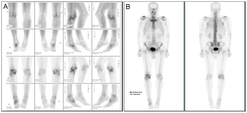
Methods: Samples of serum and knee joint synovial fluid (275 knees) were obtained from 159 patients with symptomatic OA of at least 1 knee. Bone scintigraphy using (99m)Tc-labeled methylene diphosphonate was performed, and early-phase knee scans and late-phase whole-body bone scans of 15 additional joint sites were scored semiquantitatively. To control for within-subject correlations of knee data, generalized linear modeling was used in the correlation of the bone scan scores with the COMP levels. Principal components analysis was used to explore the contribution of each joint site to the variance in serum COMP levels.
Results: The correlation between synovial fluid and serum COMP levels was significant (r = 0.206, P = 0.006). Synovial fluid COMP levels correlated most strongly with the early-phase knee bone scan scores (P = 0.0003), even after adjustment for OA severity according to the late-phase bone scan scores (P = 0.015), as well as synovial fluid volumes (P < 0.0001). Serum COMP levels correlated with the total-body bone scan scores (r = 0.188, P = 0.018) and with a factor composed of the bone scan scores in the shoulders, spine, lateral knees, and sacroiliac joints (P = 0.0004).
Conclusion: Synovial fluid COMP levels correlated strongly with 2 indicators of knee joint inflammation: early-phase bone scintigraphic findings and synovial fluid volume. Serum COMP levels correlated with total-body joint disease severity as determined by late-phase bone scintigraphy, supporting the hypothesis that whole-body bone scintigraphy is a means of quantifying the total-body burden of OA for systemic biomarker validation.
Figures
Similar articles
-
[Cartilage oligomeric matrix protein (COMP): the role of a non-collagen cartilage matrix protein as a marker of disease activity and joint destruction in patients with rheumatoid arthritis and osteoarthritis].Z Rheumatol. 1999 Apr;58(2):79-87. doi: 10.1007/s003930050156. Z Rheumatol. 1999. PMID: 10408068 German.
-
Serum cartilage oligomeric matrix protein reflects osteoarthritis presence and severity: the Johnston County Osteoarthritis Project.Arthritis Rheum. 1999 Nov;42(11):2356-64. doi: 10.1002/1529-0131(199911)42:11<2356::AID-ANR14>3.0.CO;2-R. Arthritis Rheum. 1999. PMID: 10555031
-
Cartilage oligomeric matrix protein in serum in systemic lupus erythematosus and knee osteoarthritis. Preliminary communication.Rheumatol Int. 2005 Jun;25(5):373-8. doi: 10.1007/s00296-004-0581-7. Epub 2005 Mar 1. Rheumatol Int. 2005. PMID: 15739097
-
Correlation of serum cartilage oligomeric matrix protein with knee osteoarthritis diagnosis: a meta-analysis.J Orthop Surg Res. 2018 Oct 19;13(1):262. doi: 10.1186/s13018-018-0959-y. J Orthop Surg Res. 2018. PMID: 30340615 Free PMC article.
-
Could Osteopontin be a useful biomarker in the diagnosis and severity assessment of osteoarthritis? A systematic review and meta-analysis of recent evidence.Clin Immunol. 2023 Jan;246:109187. doi: 10.1016/j.clim.2022.109187. Epub 2022 Nov 17. Clin Immunol. 2023. PMID: 36403917
Cited by
-
Multiple joint osteoarthritis (MJOA): What's in a name?Osteoarthritis Cartilage. 2024 Mar;32(3):234-240. doi: 10.1016/j.joca.2023.10.008. Epub 2023 Nov 19. Osteoarthritis Cartilage. 2024. PMID: 37984559 Free PMC article. Review.
-
Synovial Fluid Biomarkers in Knee Osteoarthritis: A Systematic Review and Quantitative Evaluation Using BIPEDs Criteria.Cartilage. 2021 Dec;13(1_suppl):82S-103S. doi: 10.1177/1947603520942941. Epub 2020 Jul 25. Cartilage. 2021. PMID: 32713185 Free PMC article.
-
The TransEurope FootRace Project: longitudinal data acquisition in a cluster randomized mobile MRI observational cohort study on 44 endurance runners at a 64-stage 4,486 km transcontinental ultramarathon.BMC Med. 2012 Jul 19;10:78. doi: 10.1186/1741-7015-10-78. BMC Med. 2012. PMID: 22812450 Free PMC article. Clinical Trial.
-
The current and future status of biomarkers in osteoarthritis.J Rheumatol. 2014 May;41(5):834-6. doi: 10.3899/jrheum.140094. J Rheumatol. 2014. PMID: 24788463 Free PMC article. No abstract available.
-
Alpha C-telopeptide of type I collagen is associated with subchondral bone turnover and predicts progression of joint space narrowing and osteophytes in osteoarthritis.Arthritis Rheumatol. 2014 Sep;66(9):2440-9. doi: 10.1002/art.38739. Arthritis Rheumatol. 2014. PMID: 24909851 Free PMC article.
References
-
- Sturmer T, Sun Y, Sauerland S, Zeissig I, Gunther KP, Puhl W, et al. Serum cholesterol and osteoarthritis. The baseline examination of the Ulm Osteoarthritis Study. J Rheumatol. 1998;25(9):1827–32. - PubMed
-
- Jonsson H, Eliasson GJ, Petursson E. Scintigraphic hand osteoarthritis (OA)--prevalence, joint distribution, and association with OA at other sites. J Rheumatol. 1999;26(7):1550–6. - PubMed
Publication types
MeSH terms
Substances
Grants and funding
LinkOut - more resources
Full Text Sources
Medical
Miscellaneous
