. 2010 Jan;51(1):329-35.
doi: 10.1002/hep.23335.

Generation of functional human hepatic endoderm from human induced pluripotent stem cells

Affiliations

Generation of functional human hepatic endoderm from human induced pluripotent stem cells

Gareth J Sullivan et al. Hepatology. 2010 Jan.

Abstract

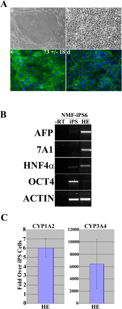
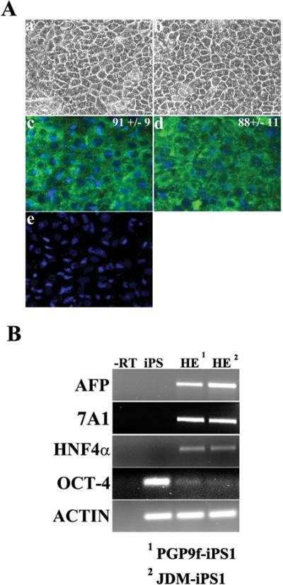
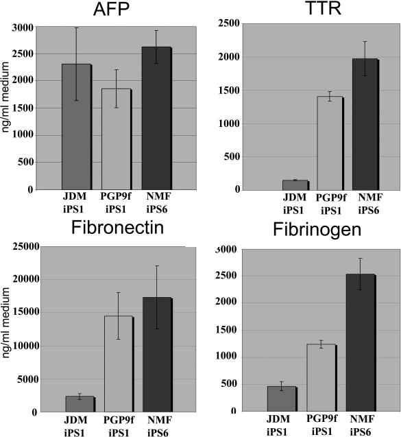
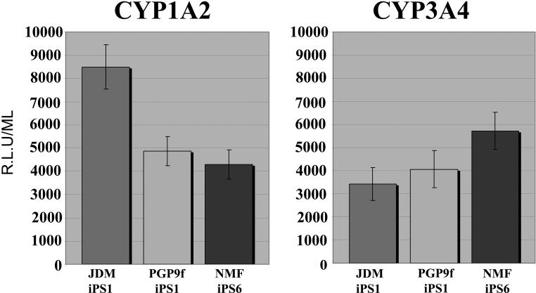
With the advent of induced pluripotent stem cell (iPSC) technology, it is now feasible to generate iPSCs with a defined genotype or disease state. When coupled with direct differentiation to a defined lineage, such as hepatic endoderm (HE), iPSCs would revolutionize the way we study human liver biology and generate efficient "off the shelf" models of human liver disease. Here, we show the "proof of concept" that iPSC lines representing both male and female sexes and two ethnic origins can be differentiated to HE at efficiencies of between 70%-90%, using a method mimicking physiological relevant condition. The iPSC-derived HE exhibited hepatic morphology and expressed the hepatic markers albumin and E-cadherin, as assessed by immunohistochemistry. They also expressed alpha-fetoprotein, hepatocyte nuclear factor-4a, and a metabolic marker, cytochrome P450 7A1 (Cyp7A1), demonstrating a definitive endodermal lineage differentiation. Furthermore, iPSC-derived hepatocytes produced and secreted the plasma proteins, fibrinogen, fibronectin, transthyretin, and alpha-fetoprotein, an essential feature for functional HE. Additionally iPSC-derived HE supported both CYP1A2 and CYP3A4 metabolism, which is essential for drug and toxicology testing.

Conclusion: This work is first to demonstrate the efficient generation of hepatic endodermal lineage from human iPSCs that exhibits key attributes of hepatocytes, and the potential application of iPSC-derived HE in studying human liver biology. In particular, iPSCs from individuals representing highly polymorphic variants in metabolic genes and different ethnic groups will provide pharmaceutical development and toxicology studies a unique opportunity to revolutionize predictive drug toxicology assays and allow the creation of in vitro hepatic disease models.


Figures

Figure 1
Figure 1. Derivation of hepatic endoderm from iPSCs
(Aa) Phase contrast microscropy demonstrating the typical iPSC colony morphology and (Ab) iPSC-derived hepatic endoderm (HE) following 14 days in the differentiation procedure (Magnification x20). (Ac) iPSC-derived HE stains positive for albumin at day 14 in the differentiation procedure. The numbers represent the efficiency of the procedure +/− SE. (Ad) iPSC-derived HE stains positive for E-Cadherin at day 14 in the differentiation procedure (B) RT-PCR gene expression of iPSCs and iPSC-derived hepatic endoderm. iPSC-derived hepatic endoderm on (lane 1) express the markers AFP, CYP7A1 and HNF4 alpha, but not OCT-4. iPSCs strongly express OCT4. Potentially due to spontaneous differentiation found in iPSC culture. PCR reactions were controlled using a –RT control (lane 4) and details of primers and cycles can be found in the supplementary methods section (C) CYP1A2 and CYP3A4.
Figure 2
Figure 2. Derivation of HE from 2 other iPSC lines
(Aa,b) Phase contrast microscropy demonstrating the typical iPSC HE morphology following 14 days in the differentiation procedure (Magnification x20). (Ac,d) IPSC-derived HE stains positive for albumin at day 14 in the differentiation procedure. The numbers represent the efficiency of the procedure +/− SE. (B) RT-PCR gene expression of iPSCs and iPSC-derived hepatic endoderm. iPSC-derived hepatic endoderm on (lane 1) express the markers AFP, and HNF4 alpha, but not OCT-4. iPSCs strongly express OCT4. Potentially due to spontaneous differentiation found in iPSC culture. PCR reactions were controlled using a –RT control (-RT) and details of primers and cycles can be found in the supplementary methods section
Figure 3
Figure 3. iPSC-derived hepatic endoderm produces hepatic specific serum proteins
iPSC-derived hepatic endoderm was maintained in 1ml of hepatocyte culture medium for 24 hours. The following morning culture supernatants were harvested and serum protein production measured by ELISA and quoted as ng/ml of tissue culture medium per 24 hours. Note background has been removed from the presented data. Each line is represented as a different coloured bar: JDM-iPS1 is presented by the first bar in all graphs, PGP9ff-iPS1 is repsresented by the middle bar in each graph and NMF-iPS6 is represented by the last bar in each graph. In all lines the serum protein tested was detected. fibrinogen and transthyretin (TTR), fibronectin and alpha-fetoprotein (AFP) (n=6).
Figure 4
Figure 4. iPSC-derived hepatic endoderm displays cytochrome P450 metabolism
iPSC-derived hepatic endoderm were incubated with hepatocyte culture medium supplemented with 50 μM of CYP3A4 or CYP1A2 pGlo TM substrates (Promega) as per manufacturers instructions. 4 hours post-treatment 50 μl of culture medium was removed and read on a luminometer (POLARstar optima). CYP1A2 and CYP3A4 activity is expressed as relative light units (R.L.U.)/ml of tissue culture medium. (n=6). CYP1A2 activity was similar on cells maintained on both ECMs. Luciferase activity is expressed as relative light units (R.L.U.)/ml of tissue culture media. JDM-iPS1 is presented by the first bar in all graphs, PGP9f-iPS1 is represented by the middle bar in each graph and NMF-iPS6 is represented by the last bar in each graph.

Comment in

References

    1. Park IH, Zhao R, West JA, Yabuuchi A, Huo H, Ince TA, et al. Reprogramming of human somatic cells to pluripotency with defined factors. Nature. 2008;451:141–146. - PubMed
    1. Takahashi K, Tanabe K, Ohnuki M, Narita M, Ichisaka T, Tomoda K, et al. Induction of pluripotent stem cells from adult human fibroblasts by defined factors. Cell. 2007;131:861–872. - PubMed
    1. Yu J, Hu K, Smuga-Otto K, Tian S, Stewart R, Slukvin II, et al. Human induced pluripotent stem cells free of vector and transgene sequences. Science. 2007;318:1917–1920. - PMC - PubMed
    1. Dalgetty DM, Medine CN, Iredale JP, Hay DC. Progress and fuure challenges in stem cell-derived liver technologies. Am. J. Physiol. Gastrointest Liver Physiol. 2009;297 doi:10.1152/ajpgi.00138.2009. - PubMed
    1. Hay DC, Fletcher J, Payne C, Terrace JD, Gallagher RC, Snoeys J, et al. Highly efficient differentiation of hESCs to functional hepatic endoderm requires ActivinA and Wnt3a signaling. Proc. Nat. Acad. Sci. 2008;105:12301–12306. - PMC - PubMed

Publication types