. 2009 Dec;238(12):3185-92.
doi: 10.1002/dvdy.22120.

Characterization of the expression pattern of the PRC2 core subunit Suz12 during embryonic development of Xenopus laevis

Affiliations

Characterization of the expression pattern of the PRC2 core subunit Suz12 during embryonic development of Xenopus laevis

Issam Aldiri et al. Dev Dyn. 2009 Dec.

Abstract

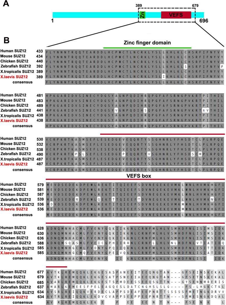
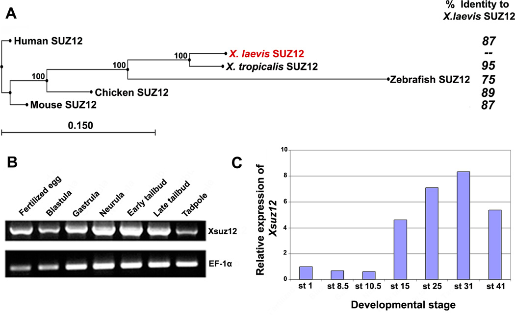
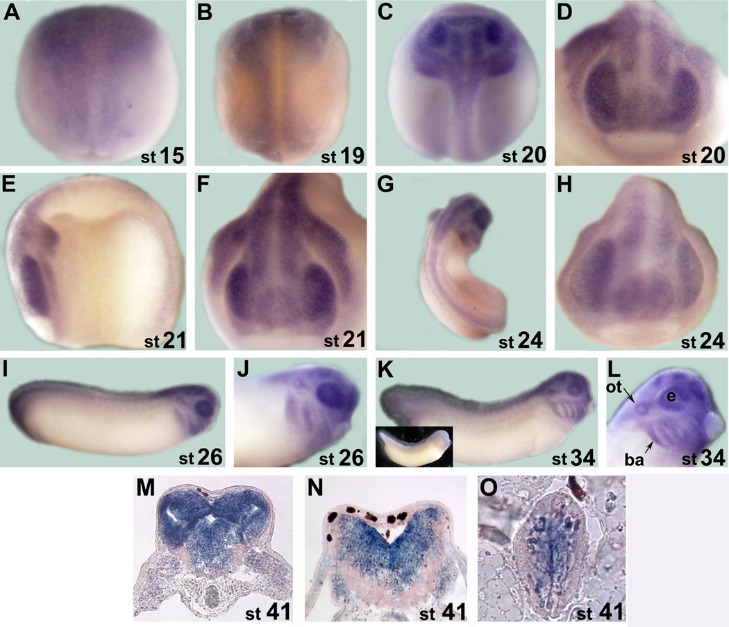
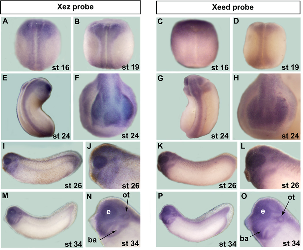
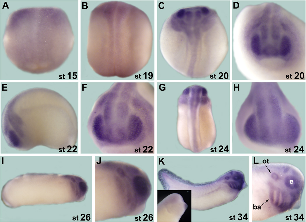
The Polycomb repressive complex 2 is a multimeric aggregate that mediates silencing of a broad range of genes, and is associated with important biological contexts such as stem cell maintenance and cancer progression. PRC2 mainly trimethylates lysine 27 of histone H3 and is composed of three essential core subunits: EZH2, EED, and SUZ12. The Xenopus orthologs of PRC2 subunits Ezh2 and Eed have been described but Suz12 remained unidentified. Here, we report the cloning of the Xenopus Suz12, and determine its spatiotemporal expression during development. Xsuz12 transcript is provided maternally and continues to be expressed throughout development, particularly in the anterior part of the developing central nervous system. Importantly, comparative analysis of the expression of the PRC2 subunits Xez, Xeed, and Xrbbp4 indicates that their expression largely coincides with Xsuz12 in the nervous system, suggesting that PRC2 may have unexplored functions in the development of the frog central nervous system.


Figures

Fig. 1
Fig. 1
Suz12 is highly conserved among vertebrates. A: schematic representation of XSUZ12 showing the relative positions of its zinc finger domain and VEFS box. B: Sequence alignment of Suz12 orthologs in vertebrates representing area boxed in A. The predicted amino acid sequence of X. laevis SUZ12 is aligned with human (Homo sapiens; accession no. NP_056170), mouse (Mus musculus; accession no. NP_954666.1), Chicken (Gallus gallus; accession no. XP_415658.), Zebrafish (Danio rerio; accession no. XP_694666) and X.tropicalis (accession no. NP_001072292). Green and red lines span the Zinc finger domain and the VEFS box, respectively.
Fig. 2
Fig. 2
A: Phylogenetic tree of SUZ12 proteins of different species created by neighbor-joining algorithm using CLC Sequence Viewer, and their respective amino acid identity compared to X. laevis Suz12. Percentage of branching point reproducibility is listed next to each node. Scale bar represents distance calculated on the basis of amino acid substitution rates. B: RT-PCR analysis of Xsuz12 expression during the frog development. cDNA pools were prepared from the following stages: Fertilized egg: stage 1, Blastula: stage 8–9, Gastrula: stage 10.5, Neurula: stage 14–18, Early tailbud: stage 25, Late tailbud; stage 35, Tadpole: stage 41. EF1-α was used as a loading control. C: Relative amounts of Xsuz12 transcripts during Xenopus development as revealed by qRT-PCR. The values of Xsuz12 was normalized to those of H4. Bars represent fold change in Xsuz12 expression at different stages as compared to stage 1.
Fig. 3
Fig. 3
A-L: Whole mount in situ hybridization analysis of Xsuz12 expression starting from stage 15 (neurula). A, B, C and G are dorsal views. D, F and H are anterior views. E, I, G, K and L are lateral views. G and L are magnifications of the head region as viewed laterally at stage 26 and 34, respectively. Inset in K shows an example of a whole mount in situ hybridization that was performed with Xsuz12 sense probe. M-O: in situ hybridization analysis performed on traverse sections from the head region at stage 41. M and N sections are at the levels of forebrain and hindbrain, respectively. O is a traverse section in the spinal cord. Stages are indicated at the lower right corner of each image. ot, otic vesicle; ba, branchial arches; e, eye.
Fig. 4
Fig. 4
Comparison of Xez and Xeed spatial expression during frog development as assessed by whole mount in situ hybridization. A B, C, D, E and G are dorsal views. F and H are anterior views. I, K, M and P are lateral views. J, L, N and O are magnifications of the head region in I, K, M and P, respectively. ot, otic vesicle; ba, branchial arches; e, eye.
Fig. 5
Fig. 5
Expression of Xrbbp4 by whole mount in situ hybridization analysis. A B, C and G are dorsal views. D, F and H are anterior views. I, K, M and P are lateral views. Whole mount in situ hybridization performed with a sense probe is shown as inset in 3K. ot, otic vesicle; ba, branchial arches; e, eye.

References

    1. Barnett MW, Seville RA, Nijjar S, Old RW, Jones EA. Xenopus Enhancer of Zeste (XEZ); an anteriorly restricted polycomb gene with a role in neural patterning. Mech Dev. 2001;102:157–167. - PubMed
    1. Barski A, Cuddapah S, Cui K, Roh TY, Schones DE, Wang Z, Wei G, Chepelev I, Zhao K. High-resolution profiling of histone methylations in the human genome. Cell. 2007;129:823–837. - PubMed
    1. Boyer LA, Plath K, Zeitlinger J, Brambrink T, Medeiros LA, Lee TI, Levine SS, Wernig M, Tajonar A, Ray MK, Bell GW, Otte AP, Vidal M, Gifford DK, Young RA, Jaenisch R. Polycomb complexes repress developmental regulators in murine embryonic stem cells. Nature. 2006;441:349–353. - PubMed
    1. Cao R, Zhang Y. SUZ12 is required for both the histone methyltransferase activity and the silencing function of the EED—EZH2 complex. Mol Cell. 2004;15:57–67. - PubMed
    1. Caretti G, Di Padova M, Micales B, Lyons GE, Sartorelli V. The Polycomb Ezh2 methyltransferase regulates muscle gene expression and skeletal muscle differentiation. Genes Dev. 2004;18:2627–2638. - PMC - PubMed

Publication types

MeSH terms

LinkOut - more resources