

Review
doi: 10.1111/j.1742-4658.2009.07401.x.
Viral entry mechanisms: the increasing diversity of paramyxovirus entry
Affiliations
- PMID: 19878307
- PMCID: PMC2795005
- DOI: 10.1111/j.1742-4658.2009.07401.x
Item in Clipboard
Review
Viral entry mechanisms: the increasing diversity of paramyxovirus entry
FEBS J.
2009 Dec.
Abstract
The paramyxovirus family contains established human pathogens such as the measles virus and human respiratory syncytial virus, as well as emerging pathogens including the Hendra and Nipah viruses and the recently identified human metapneumovirus. Two major envelope glycoproteins, the attachment protein and the fusion protein, promote the processes of viral attachment and virus-cell membrane fusion required for entry. Although common mechanisms of fusion protein proteolytic activation and the mechanism of membrane fusion promotion have been shown in recent years, considerable diversity exists in the family relating to receptor binding and the potential mechanisms of fusion triggering.
Figures
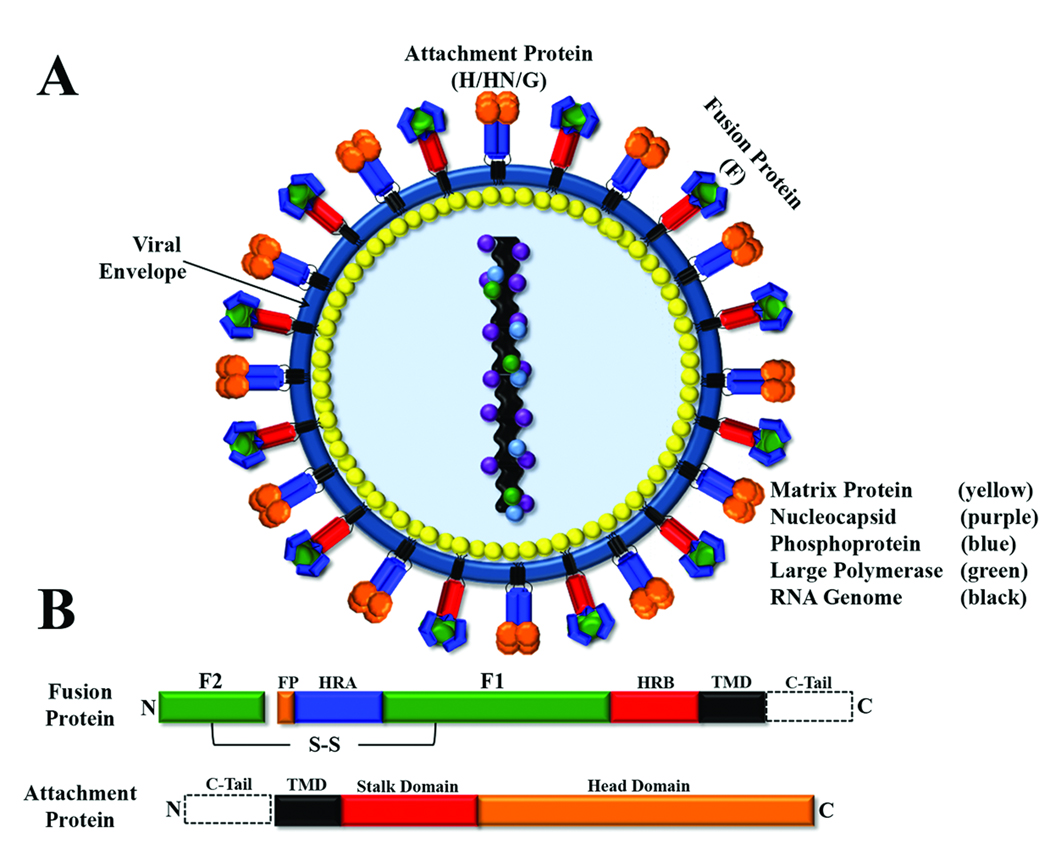
A) Schematic of a paramyxovirus; viral membrane shown in blue. B) Conserved domains of paramyxovirus fusion and attachment proteins. Domain abbreviations: fusion peptide (FP, orange); heptad repeat A (HRA, blue); heptad repeat B (HRB, red); transmembrane domain (TMD, black); cytoplasmic tail (C-Tail, dotted box); disulfide bond (S-S).
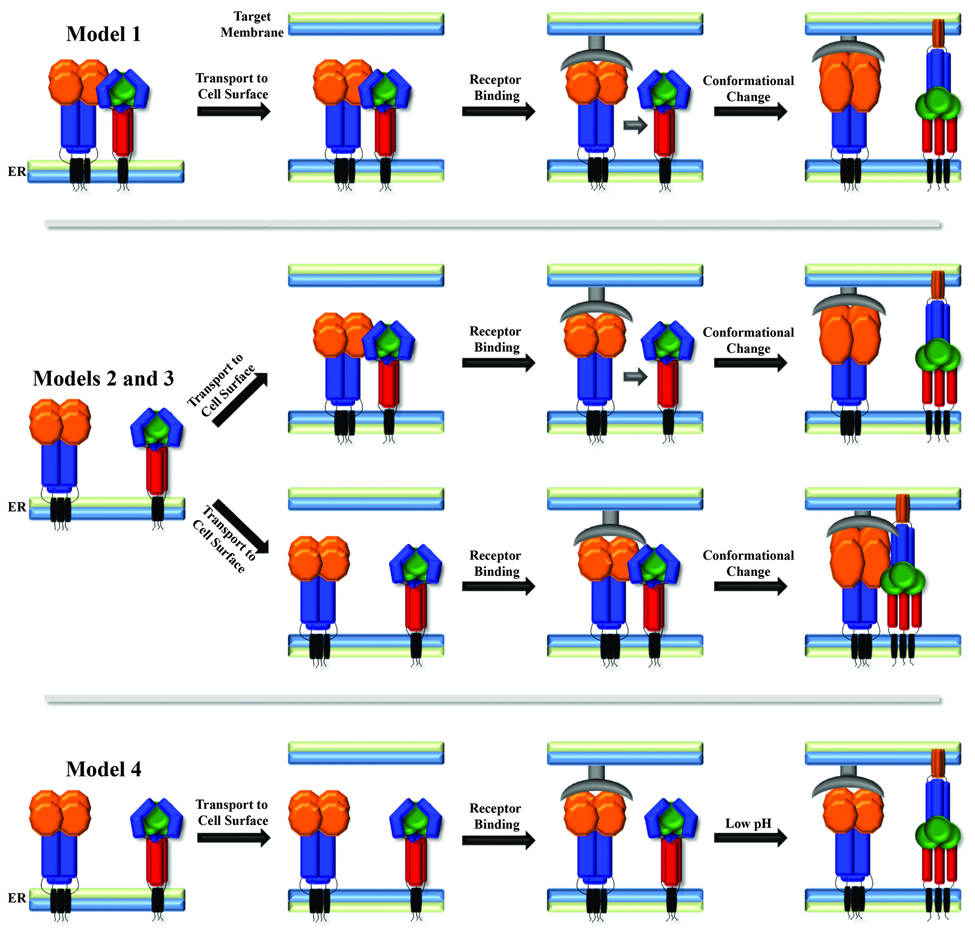
Attachment protein shown with orange head domain and blue stalk, fusion protein shown in blue/green head domain and red stalk region, receptor shown in grey.

A) Lipid intermediates culminating in the formation of a full fusion pore. B) Proposed fusion protein intermediates with subsequent formation of the post-fusion six-helix bundle. Fusion peptide,orange; heptad repeat A, blue; heptad repeat B, red; transmembrane domain, black.
References
-
- Lamb RA, Parks GD. Paramyxoviridae: the viruses and their replication. In: Knipe DM, Howley PM, editors. Fields Virology. Lippincott: Williams and Wilkins; 2007. pp. 1449–1496.
-
- Tong S, Compans RW. Alternative mechanisms of interaction between homotypic and heterotypic parainfluenza virus HN and F proteins. J. Gen. Virol. 1999;80:107–115. - PubMed
Publication types
MeSH terms
Substances
Grants and funding
LinkOut - more resources
Full Text Sources
Other Literature Sources
