

Cleavage of functional IL-2 receptor alpha chain (CD25) from murine corneal and conjunctival epithelia by MMP-9
- PMID: 19878594
- PMCID: PMC2777897
- DOI: 10.1186/1476-9255-6-31
Cleavage of functional IL-2 receptor alpha chain (CD25) from murine corneal and conjunctival epithelia by MMP-9
Abstract
Background: IL-2 has classically been considered a cytokine that regulates T cell proliferation and differentiation, signaling through its heterotrimeric receptor (IL-2R) consisting of alpha (CD25), beta (CD122), gamma chains (CD132). Expression of IL-2R has also been detected in mucosal epithelial cells. Soluble IL-2Ralpha (CD25) has been reported as an inflammatory marker. We evaluated the expression of CD25 and CD122 in the ocular surface epithelium and investigated the mechanism of proteolytic cleavage of CD25 from these cells.
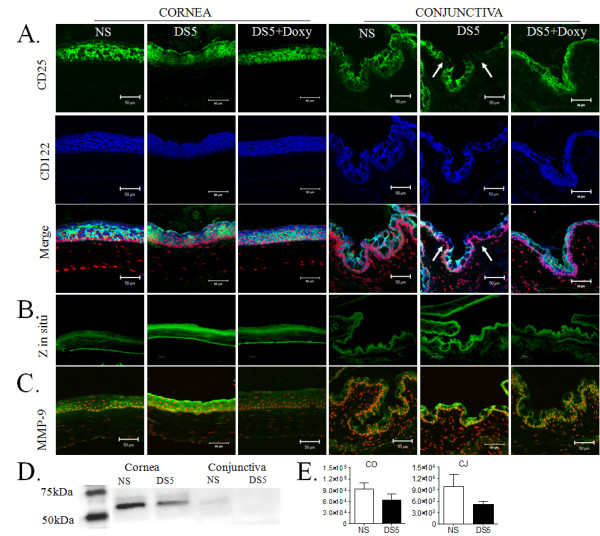
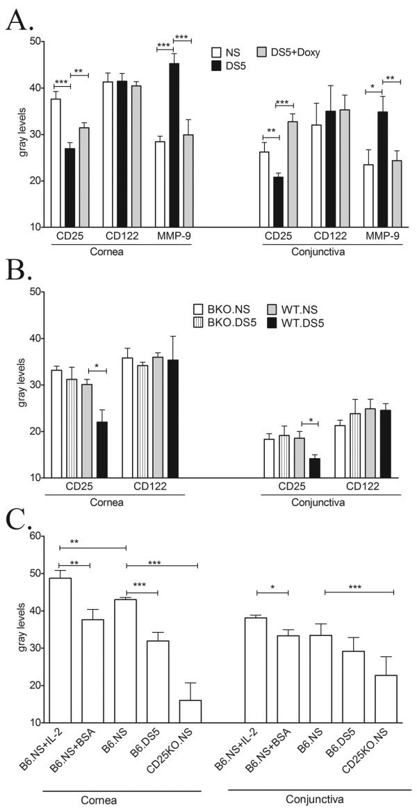
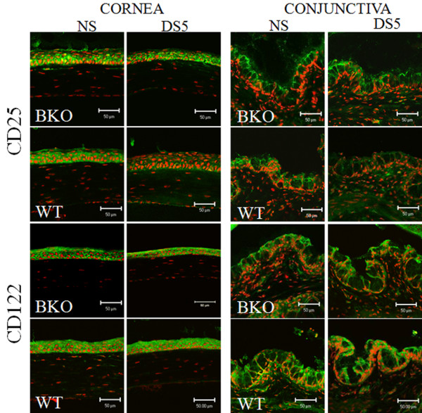
Methods: Desiccating stress (DS) was used as an inducer of matrix metalloproteinase 9 (MMP-9). DS was created by subjecting C57BL/6 and MMP-9 knockout (BKO) mice and their wild-type littermates (WT) mice to a low humidity and drafty environment for 5 days (DS5). A separate group of C57BL/6 mice was subjected to DS5 and treatment with topical 0.025% doxycycline, a MMP inhibitor, administered QID. The expression of CD25 and CD122 was evaluated in cryosections by dual-label laser scanning confocal microscopy. Western blot was used to measure relative levels of CD25 in epithelial lysates. Gelatinase activity was evaluated by in situ zymography. Soluble CD25 in tear fluid was measured by an immunobead assay.
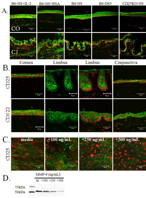
Results: CD25 and CD122 were abundantly expressed in cornea (all layers) and conjunctiva epithelia (apical and subapical layers) in nonstressed control mice. After desiccating stress, we found that immunoreactivity to CD25, but not CD122, decreased by the ocular surface epithelia and concentration of soluble CD25 in tears increased as MMP-9 staining increased. CD25 was preserved in C57BL/6 mice topically treated with an MMP-9 inhibitor and in MMP-9 knock-out mice. MMP-9 treatment of human cultured corneal epithelial cells decreased levels of CD25 protein in a concentration dependent fashion.
Conclusion: Our results indicate that functional IL-2R is produced by the ocular surface epithelia and that CD25 is proteolytic cleaved to its soluble form by MMP-9, which increases in desiccating stress. These findings provide new insight into IL-2 signaling in mucosal epithelia.
Figures
Similar articles
-
Corticosteroid and doxycycline suppress MMP-9 and inflammatory cytokine expression, MAPK activation in the corneal epithelium in experimental dry eye.Exp Eye Res. 2006 Sep;83(3):526-35. doi: 10.1016/j.exer.2006.02.004. Epub 2006 Apr 27. Exp Eye Res. 2006. PMID: 16643899
-
Conditional deletion of CD25 in the corneal epithelium reveals sex differences in barrier disruption.Ocul Surf. 2023 Oct;30:57-72. doi: 10.1016/j.jtos.2023.07.008. Epub 2023 Jul 27. Ocul Surf. 2023. PMID: 37516317 Free PMC article.
-
Experimental dry eye stimulates production of inflammatory cytokines and MMP-9 and activates MAPK signaling pathways on the ocular surface.Invest Ophthalmol Vis Sci. 2004 Dec;45(12):4293-301. doi: 10.1167/iovs.03-1145. Invest Ophthalmol Vis Sci. 2004. PMID: 15557435
-
Clusterin in the eye: An old dog with new tricks at the ocular surface.Exp Eye Res. 2016 Jun;147:57-71. doi: 10.1016/j.exer.2016.04.019. Epub 2016 Apr 27. Exp Eye Res. 2016. PMID: 27131907 Review.
-
The IL-2A receptor pathway and its role in lymphocyte differentiation and function.Cytokine Growth Factor Rev. 2022 Oct;67:66-79. doi: 10.1016/j.cytogfr.2022.06.004. Epub 2022 Jul 2. Cytokine Growth Factor Rev. 2022. PMID: 35803834 Review.
Cited by
-
Anti-CCR4 monoclonal antibody enhances antitumor immunity by modulating tumor-infiltrating Tregs in an ovarian cancer xenograft humanized mouse model.Oncoimmunology. 2015 Dec 10;5(3):e1090075. doi: 10.1080/2162402X.2015.1090075. eCollection 2016 Mar. Oncoimmunology. 2015. PMID: 27141347 Free PMC article.
-
IL-22 from conventional NK cells is epithelial regenerative and inflammation protective during influenza infection.Mucosal Immunol. 2013 Jan;6(1):69-82. doi: 10.1038/mi.2012.49. Epub 2012 Jun 27. Mucosal Immunol. 2013. PMID: 22739232 Free PMC article.
-
EMMPRIN modulates epithelial barrier function through a MMP-mediated occludin cleavage: implications in dry eye disease.Am J Pathol. 2011 Sep;179(3):1278-86. doi: 10.1016/j.ajpath.2011.05.036. Epub 2011 Jul 21. Am J Pathol. 2011. PMID: 21777561 Free PMC article.
-
Clinical significance of sIL-2R levels in B-cell lymphomas.PLoS One. 2013 Nov 13;8(11):e78730. doi: 10.1371/journal.pone.0078730. eCollection 2013. PLoS One. 2013. PMID: 24236041 Free PMC article.
-
Roles for trafficking and O-linked glycosylation in the turnover of model cell surface proteins.J Biol Chem. 2014 Jul 11;289(28):19477-90. doi: 10.1074/jbc.M114.564666. Epub 2014 Jun 2. J Biol Chem. 2014. PMID: 24891503 Free PMC article.
References
-
- Waldmann TA, Dubois S, Tagaya Y. Contrasting roles of IL-2 and IL-15 in the life and death of lymphocytes: implications for immunotherapy. Immunity. 2001;14:105–110. - PubMed
Grants and funding
LinkOut - more resources
Full Text Sources
Other Literature Sources
Miscellaneous
