. 2009 Dec 25;390(4):1149-53.
doi: 10.1016/j.bbrc.2009.10.106. Epub 2009 Oct 28.

Light-dependent phosphorylation of the gamma subunit of cGMP-phophodiesterase (PDE6gamma) at residue threonine 22 in intact photoreceptor neurons

Affiliations

Light-dependent phosphorylation of the gamma subunit of cGMP-phophodiesterase (PDE6gamma) at residue threonine 22 in intact photoreceptor neurons

Kerstin M Janisch et al. Biochem Biophys Res Commun. .

Abstract

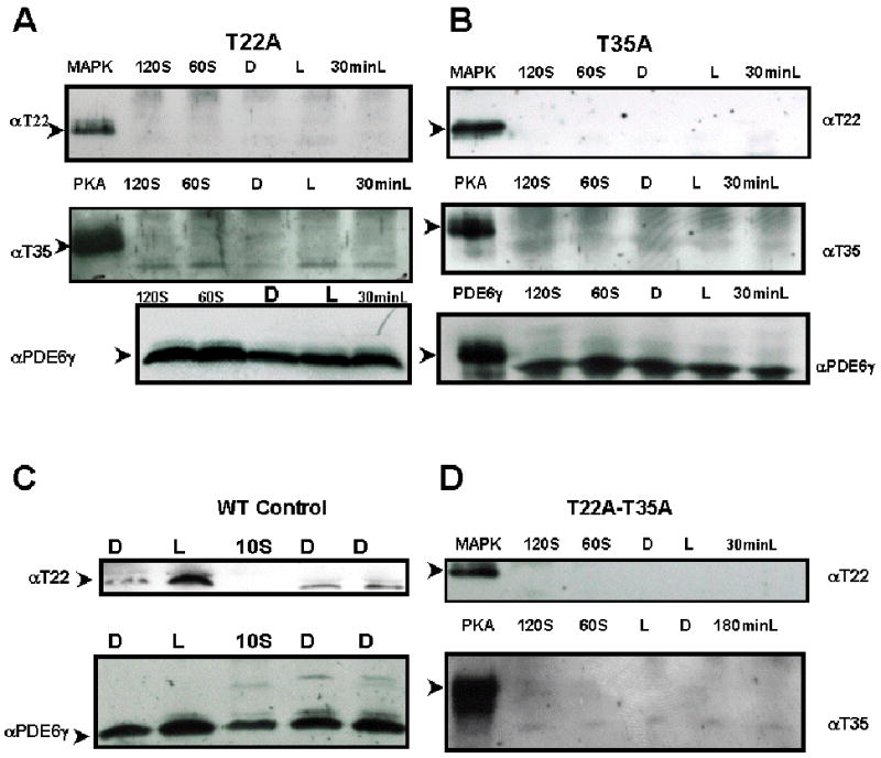
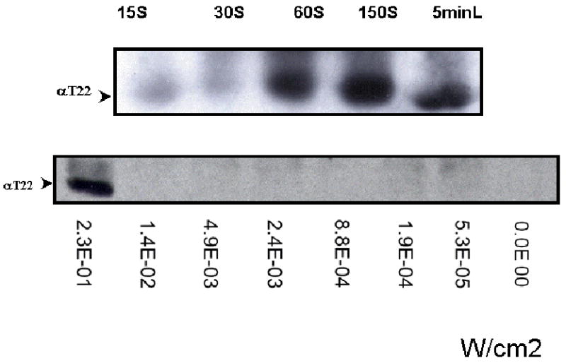
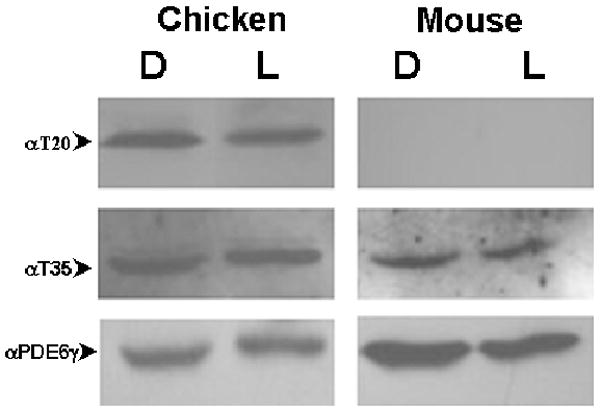
The gamma subunit of rod-specific cGMP phosphodiesterase 6 (PDE6gamma), an effector of the G-protein GNAT1, is a key regulator of phototransduction. The results of several in vitro biochemical reconstitution experiments conducted to examine the effects of phosphorylation of PDE6gamma on its ability to regulate the PDE6 catalytic core have been inconsistent, showing that phosphorylation of PDE6gamma may increase or decrease the ability of PDE6gamma to deactivate phototransduction. To resolve role of phosphorylation of PDE6gamma in living photoreceptors, we generated transgenic mice in which either one or both Threonine (T) sites in PDE6gamma (T22 and T35), which are candidates for putative regulatory phosphorylation, were substituted with alanine (A). Phosphorylation of these sites was examined as a function of light exposure. We found that phosphorylation of T22 increases with light exposure in intact mouse rods while constitutive phosphorylation of T35 is unaffected by light in intact mouse rods and cones. Phosphorylation of the cone isoform of PDE6gamma, PDE6H, is constitutively phosphorylated at the T20 residue. Light-induced T22 phosphorylation was lost in T35A transgenic rods, and T35 phosphorylation was extinguished in T22A transgenic rods. The interdependency of phosphorylation of T22 and T35 suggests that light-induced, post-translational modification of PDE6gamma is essential for the regulation of G-protein signaling.


Figures

Fig. 1
Fig. 1
Both anti-pT22 and anti-pT35 antibodies recognized specific residues in phosphorylated PDE6γ. Immunoblot of mutant transgenic retinal extracts probed with rabbit anti-phosphoT22 antibody recognizing the phosphorylated T22 residue of PDE6γ or rabbit anti-phosphoT35 antibody recognizing the phosphorylated T35 residue of PDE6. MAPK, mitogen-activated protein kinase phosphorylated PDE6γ marked the expected molecular weight of pT22- PDE6γ at 11 kDa (top row, Fig 1A, B and D). PKA, protein kinase A phosphorylated PDE6γ marked the expected molecular weight of pT35- PDE6γ (second row, Fig 1A, B and D). Panel A: T22A transgenic retinal extract. T35 phosphorylation was lost in T22A transgenic rods. Panel B: T35A transgenic retinal extract. Light induced T22 phosphorylation is lost in T35A transgenic rods. Panel C: Control wild-type MF1 retinal extract. Light induced T22 phosphorylation was seen. Panel D: T22A-T35A transgenic retinal extract. Both light-induced T22 and basal T35 phosphorylation was significantly diminished. Presence of PDE6γ in each sample was confirmed by stripping and re-staining the filter with rabbit anti-native PDE6γ antibody. Reprobing with anti-PDE6γ antibody demonstrated expression of PDE6γ protein in these lysates. D, overnight dark-adapted retinal extracts; L, light-adapted; 60S, 60 seconds of light exposure to a dark-adapted retina; 120S, 120 seconds of light exposure to a dark-adapted retina; 30minL, 30 minutes of light exposure to a dark-adapted retina; 180 minL, 180 minutes of light exposure to a dark-adapted retina. MAPK, mitogen-activated protein kinase phosphorylated PDE6; PKA, protein kinase A phosphorylated PDE6. Both pT22 and pT35 antibodies only recognized the phosphorylated cognate peptides and anti-pT22 only recognized MAP kinase-treated (phosphorylates T22), while anti-pT35 only recognized PKA kinase-treated (phosphorylates T35) recombinant PDE6γ.
Fig. 2
Fig. 2
Linearity between T22 PDE6γ phosphorylation and Light Intensities. Panel A: T22 phosphorylation occurs within one minute of illumination. Retinas were removed and placed in a Petri dish with 5 μl of Ringer buffer to prevent drying-out. The Petri dish was placed under a light source and the dark-adapted retinas were exposed to different irradiances: 2.48× 10⁻², 4.93×10⁻², 8.63×10⁻², 2.33×10⁻¹, 4.73×10⁻¹ (W/cm2) for 15S 30S, 60S, 150S and 5 minutes (5minL), respectively. Panel B: the dark-adapted retinas were exposed to a single flash of increasing intensities: 5.3× 10⁻⁵, 1.9× 10⁻⁴, 8.8× 10⁻⁴, 2.4× 10⁻³, 1.4× 10 , and 2.3× 10⁻¹ W/cm2. T22 phosphorylation was not detected until the flash increased to 2.3× 10⁻¹ W/cm2. The irradiance (W/cm2) of the light source was determined with a light meter (Research Radiometer IL1700, International Light Inc., MA) containing a sensor fitted with a photopic filter (Y#23654).
Fig. 3
Fig. 3
PDE6H phosphorylation in chicken and murine retina. Immunoblot of normalized dark- and light-adapted chicken and murine retinal extracts probed with rabbit anti-phosphoT20 {TTGDAPTGPT(pT)PR} antibody recognizing the phosphorylated T20 residue of PDE6H or rabbit anti-pT35 antibody recognizing the phosphorylated T35 residue of PDE6γ. Phosphorylated T20 was not detected in mouse retinal extract. Presence of PDE6γ in each sample was confirmed by stripping and re-staining the filter with rabbit anti-native PDE6γ antibody. D, dark-adapted overnight; L, light-adapted to 1.67×10⁻¹ W/cm2 for sixteen hours.

References

    1. Tsang SH, Burns ME, Calvert PD, Gouras P, Baylor DA, Goff SP, Arshavsky VY. Role of the Target Enzyme in Deactivation of Photoreceptor G Protein in Vivo. Science. 1998;282:117–121. - PubMed
    1. Tsang SH, Woodruff ML, Chen CK, Yamashita CY, Cilluffo MC, Rao AL, Farber DB, Fain GL. GAP-independent termination of photoreceptor light response by excess gamma subunit of the cGMP-phosphodiesterase. J Neurosci. 2006;26:4472–80. - PMC - PubMed
    1. Tsang SH, Gouras P. Molecular Physiology and Pathology of the Retina. In: Tasman W, Jaeger EA, editors. Duane's Clinical Opthalmology. J.B. Lippincott; Philadelphia: 1996. Chapter 2.
    1. Tsang SH, Gouras P. Photoreceptors and photoreceptor dysfunctions. In: Adelman G, Smith B, editors. Encyclopedia of Neuroscience. Elsevier Science; Amsterdam: 2004. pp. 1633–1644.
    1. Tsang SH, Yamashita CK, Doi K, Salchow DJ, Bouvier N, Mendelsohn M, Gouras P, Farber DB, Goff SP. In vivo studies of the gamma subunit of retinal cGMP-phophodiesterase with a substitution of tyrosine-84. Biochem J. 2001;353:467–74. - PMC - PubMed

MeSH terms