. 2009 Nov;148(5):733-743.e2.
doi: 10.1016/j.ajo.2009.06.028.

Reticular macular disease

Affiliations

Reticular macular disease

R Theodore Smith et al. Am J Ophthalmol. 2009 Nov.

Abstract

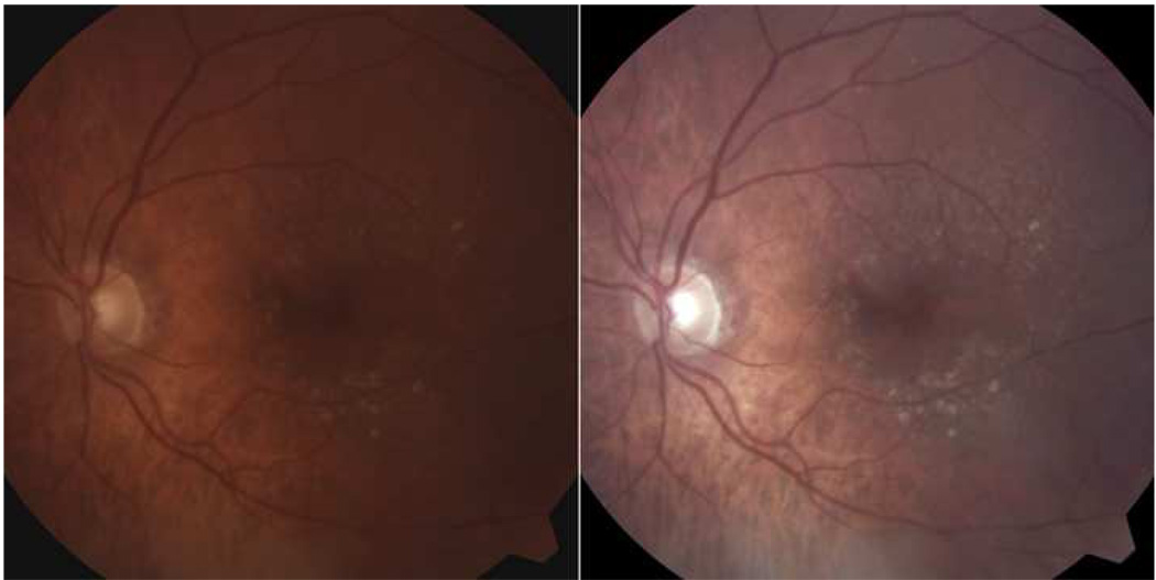
Purpose: To present a unified description of reticular macular disease (RMD), a common clinical entity that includes reticular pseudodrusen (RPD) and confers high-risk of progression to advanced age-related macular degeneration.

Design: Population-based, retrospective, cross-sectional study. Forty-two patients with reticular findings in at least one imaging method, of whom 21 were followed up.

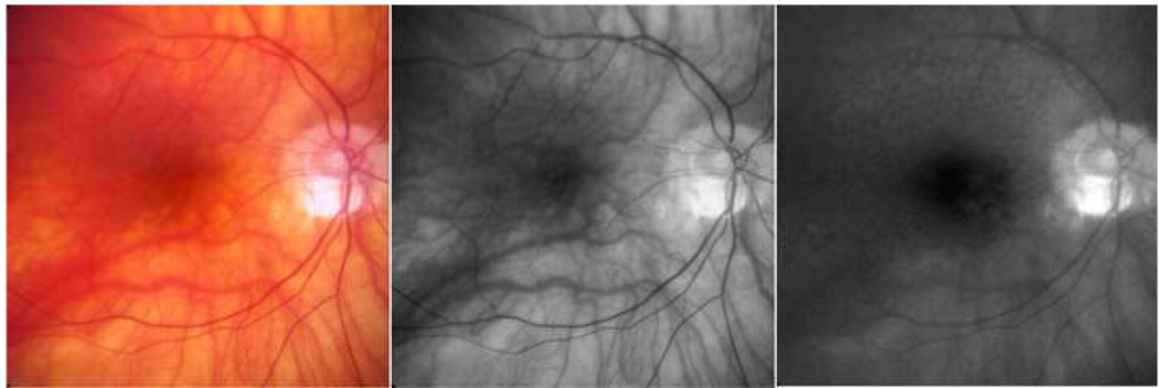
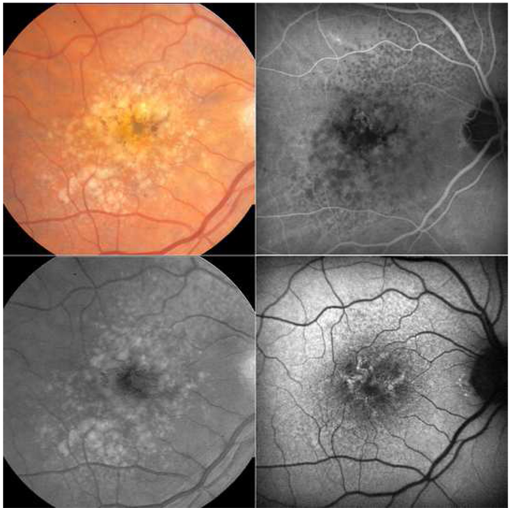
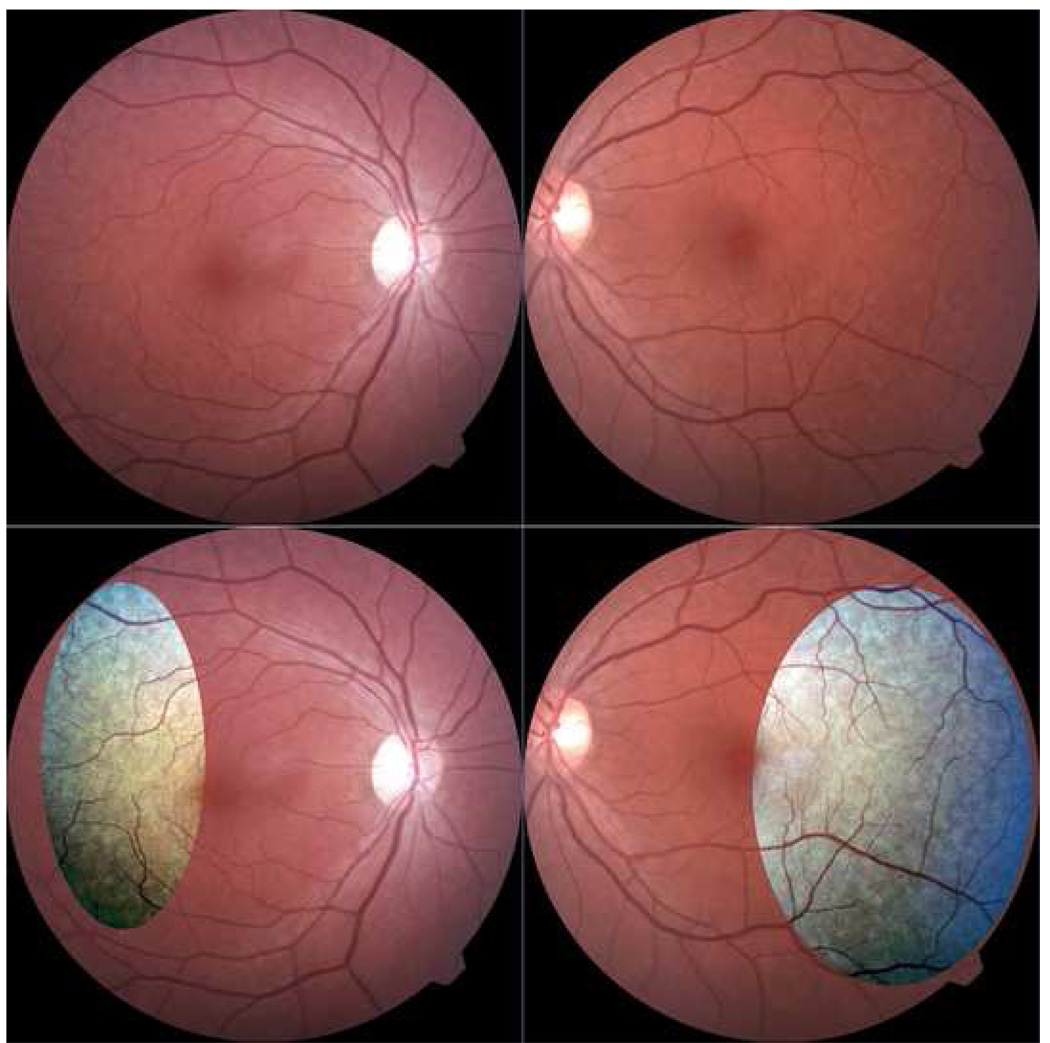
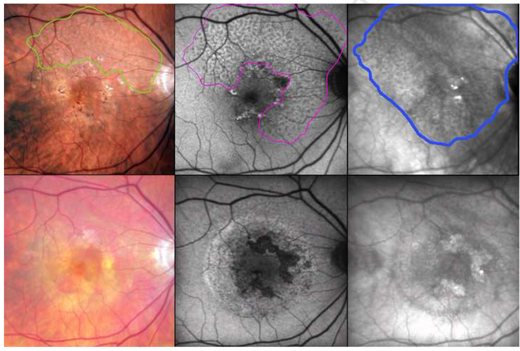
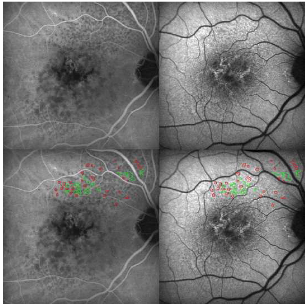
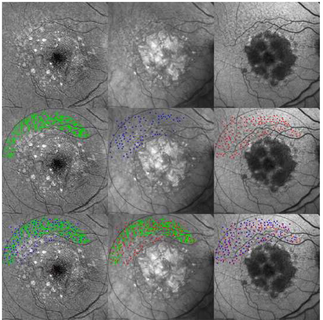
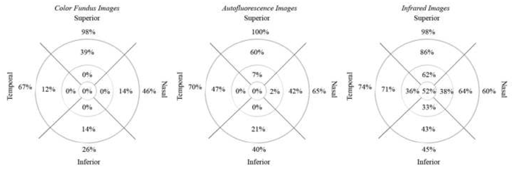
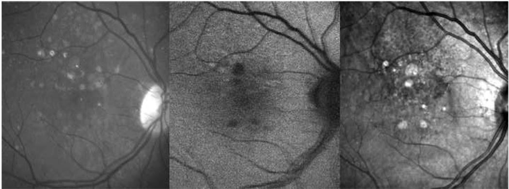
Methods: RMD was defined as RPD in color or red-free photography, in a reticular pattern on scanning laser ophthalmoscope imaging (autofluorescence scans, infrared photographs, or indocyanine green angiography), or both. Color and red-free images were contrast-enhanced, and color photographs were examined in green and blue channels. Image registration in different methods allowed comparison of areas involved and assessment of lesion colocalization.

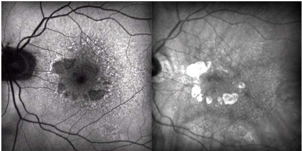
Results: RMD generally was present in both photography and scanning laser ophthalmoscope imaging. When present in two image methods, areas of RMD either largely overlapped or one fell within the other. Individual lesions had high spatial correspondence. Serial imaging showed faded to absent findings in eyes in which choroidal neovascularization developed.

Conclusions: RMD is a single disease entity with stereotypical presentations in multiple imaging methods, of which RPD is one. Autofluorescence, infrared imaging, and indocyanine green angiography suggest that it involves the retinal pigment epithelium and choriocapillaris, whereas photographic patterns implicate the inner choroid. Infrared imaging, unlike other methods, can demonstrate RMD in the central macula. RMD is associated with progression to advanced age-related macular degeneration, perhaps on an inflammatory basis. RMD deserves wider recognition among clinicians caring for elderly patients.


Figures

Figure 1
Figure 1. Color image of patient with reticular macular disease before and after contrast-enhancement
(Left) Color photo of the left eye showing large soft drusen centrally. Careful inspection reveals reticular pseudodrusen (RPD) in the superior macula. (Right) Contrast-enhanced version of image on left. The RPD in a uniform, interlacing pattern are easily seen.
Figure 2
Figure 2. Blue-channel visualization of reticular pseudodrusen (RPD) in a patient with reticular macular disease
The contrast-enhanced color photo (left) and red-free photo (center) are negative for RPD. However, the blue channel of the contrast-enhanced color photo (right) shows a definite reticular pattern superiorly.
Figure 3
Figure 3. Overall spatial correspondence of reticular lesions between imaging types, characteristic of reticular macular disease
(Top left). Enhanced color photo with reticular pseudodrusen superiorly. (Top right) Blue channel of color photo shows a reticular pattern superiorly. (Bottom left) Mid-phase indocyanine green (ICG) angiography showing a reticular pattern extending from the superior macula to the arcade and above the disc. (Bottom right) Autofluorescence scan with reticular pattern in exactly the same distribution as the patterns on the ICG and color images.
Figure 4
Figure 4. Spatial correspondence between autofluorescence (AF) and infrared (IR) reticular patterns, characteristic of reticular macular disease
(Left) AF scan of the left eye (OS) showing reticular pattern temporal and superiorly to the more typical AF lesions of age-related macular degeneration (dark areas of impending atrophy and hyper-AF overlying soft drusen). (Right) IR scan OS showing a reticular pattern of hypo-reflectant lesions with hyper-reflectant borders mostly temporally and falling within the areas of reticular AF.
Figure 5
Figure 5. Reticular pseudodrusen without maculopathy in a patient with reticular macular disease
Contrast-enhanced color photos. This 50-year-old female has significant reticular pseudodrusen peripherally bilaterally in the absence of other age-related maculopathy. The pale spot centrally in the right eye is artifact.
Figure 6
Figure 6. Fading reticular patterns across imaging modalities after choroidal neovascularization, following the pattern seen in patients with reticular macular disease
The contrast-enhanced color photo shows reticular pseudodrusen (RPD) in the superior quadrants, outlined in green (top left). The autofluorescence (AF) scan, registered to the color photo, shows a reticular pattern in 3 quadrants, outlined in magenta (top center). The infrared (IR) scan, also registered, shows a reticular pattern in all 4 quadrants extending through the central macula, outlined in blue (top right). The area involved on IR includes that involved on AF, which in turn includes the area of RPD. (Bottom row) Follow-up images were obtained after two years, during which time choroidal neovascularization developed. The enhanced color photo no longer shows RPD (bottom left), nor did the red-free or the blue channel photos (not shown). The reticular AF and IR patterns are faint, even in the areas not involved by exudation (bottom center and right, respectively).
Figure 7
Figure 7. Marked spatial correspondence between reticular lesions on autofluorescence and indocyanine green angiography, characteristic of reticular macular disease
Late-phase indocyanine green angiography (ICG, top left) and autofluorescence (AF, top right) images for the same patient are shown. Tracings of reticular pattern on the ICG photograph (red) and the autofluorescence photograph (green) were done on a separate layer using the pencil tool in Photoshop. The mask was then super-imposed onto the same ICG (bottom left) and AF (bottom right) photographs. Tracings correspond to reticular findings on both photographs, regardless of the image used to create the outlines.
Figure 8
Figure 8. Marked spatial correspondence between reticular pseudodrusen (RPD) and reticular findings on autofluorescence (AF) and infrared (IR) images, characteristic of reticular macular disease
Reticular lesions extend peripherally with visible geographic atrophy and soft drusen centrally in the contrast-enhanced blue channel color fundus photo (top left), IR (top center), and AF (top right) images of the same eye, all of which were registered and cropped. (Center left) The automated drusen segmentation tool applied in the 6000-micron region created a segmentation mask for RPD (green). (Center middle) IR reticular lesions manually marked as dots (blue) (Center right) AF reticular lesions manually marked as dots (red). These segmentations in turn are superimposed pairwise to give: (Bottom left) IR lesions (blue) overlaid on RPD (green). (Bottom center) AF lesions (red) overlaid on RPD (green). (Bottom right) AF lesions (red) overlaid on IR lesions (blue). The final results show striking co-localizations between the reticular lesions from each image modality.
Figure 9
Figure 9. Percent distributions of reticular lesions in different imaging modalities for study patients with reticular macular disease
Percent distributions of reticular pattern, when positive, in color fundus, autofluorescence, and infrared images, displayed from left to right, respectively. The reticular pattern was most frequently noted in the outer macula and outside of the grid, near the arcades, and was notably absent in the inner and central macula of color fundus images, likely secondary to pre-existing macular pathology from moderate-to-late stage age-related macular degeneration.
Figure 10
Figure 10. Negative reticular findings in color and autofluorescence (AF) images with positive findings on infrared (IR) imaging in a patient with reticular macular disease
Green-channel color fundus photograph (left), AF (center), and IR (right) images are shown. Color and AF images were graded negative for reticular findings, but IR shows a clear reticular pattern supero-nasally.

References

    1. Mimoun G, Soubrane G, Coscas G. Macular Drusen. J Fr Ophtalmol. 1990;13:511–530. Medline. - PubMed
    1. Klein R, Davis MD, Magli YL, Segal P, Klein BEK, Hubbard L. The Wisconsin Age-Related Maculopathy Grading System. Ophthalmology. 1991;98:1128–1134. Medline. - PubMed
    1. Arnold JJ, Sarks SH, Killingsworth MC, Sarks JP. Reticular Pseudodrusen: A Risk Factor in Age-Related Maculopathy. Retina. 1995;15:183–191. Medline. doi:10.1097/00006982-199515030-00001. - PubMed
    1. Cohen SY, Dubois L, Tadayoni R, Delahaye-Mazza C, Debibie C, Quentel G. Prevalence of Reticular Pseudodrusen in Age-Related Macular Degeneration with Newly Diagnosed Choroidal Neovascularization. Br J Ophthalmol. 2007;91:354–359. Medline. doi:10.1136/bjo.2006.101022. - PMC - PubMed
    1. Klein R, Meuer SM, Knudtson MD, Iyengar SK, Klein BEK. The Epidemiology of Retinal Reticular Drusen. Am J Ophthalmol. 2008;145:317–326. Medline. doi:10.1016/j.ajo.2007.09.008. - PMC - PubMed

Publication types