. 2009 Dec 1;19(22):1937-42.
doi: 10.1016/j.cub.2009.09.055. Epub 2009 Oct 29.

Deviant kinetochore microtubule dynamics underlie chromosomal instability

Affiliations

Deviant kinetochore microtubule dynamics underlie chromosomal instability

Samuel F Bakhoum et al. Curr Biol. .

Abstract

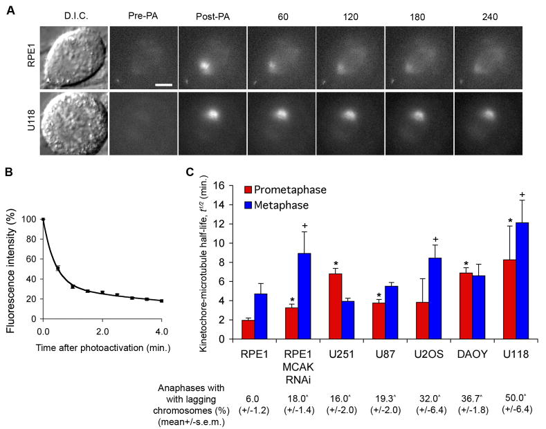
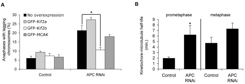
The persistent malattachment of microtubules to chromosomes at kinetochores is a major mechanism of chromosomal instability (CIN) [1, 2]. In normal diploid cells, malattachments arise spontaneously and are efficiently corrected to preserve genomic stability [3]. However, it is unknown whether cancer cells with CIN possess the ability to efficiently correct attachment errors. Here we show that kinetochore microtubule attachments in cancer cells with CIN are inherently more stable than those in normal diploid RPE-1 cells. The observed differences in attachment stability account for the persistence of malattachments into anaphase, where they cause chromosome missegregation. Furthermore, increasing the stability of kinetochore microtubule attachments in normal diploid RPE-1 cells, either by depleting the tumor suppressor protein APC or the kinesin-13 protein MCAK, is sufficient to promote chromosome segregation defects to levels comparable to those in cancer cells with CIN. Collectively, these data identify that cancer cells have a diminished capacity to correct erroneous kinetochore microtubule attachments and account for the widespread occurrence of CIN in tumors [4].


Figures

Figure 1
Figure 1. Deviant kinetochore-microtubule dynamics in cancer cells
(A) Examples of D.I.C. and time-lapse fluorescent images of spindles of RPE-1 and U118 cells before (Pre-PA) and at the indicated times (s) after activation (Post-PA) of GFP-tubulin fluorescence. Scale bar, 5 μm. (B) Example of normalized fluorescence intensity over time after photoactivating spindles of RPE1 cells at metaphase. Datapoints represent mean ± s.e.m., n = 11 cells. (C) Kinetochore-microtubule half-life (min.) in cancer cell lines at prometaphase (blue) and metaphase (red). Percent of cells at anaphase with lagging chromosomes for each cell line is denoted below the x-axis. Error bars in the graph represent standard-error derived from the exponential decay curve of the photoactivated fluorescence, (r2>0.99). *p < 0.05, †p<0.05, t-test, when compared to RPE-1 values at prometaphase and metaphase, respectively, n = 9–19 cells. ^p < 0.05, t-test, when compared to control RPE-1 values, n = 150 cells, 3 experiments.
Figure 2
Figure 2. APC loss leads to hyperstable kinetochore-microtubule attachments
(A) Percent of cells at anaphase with lagging chromosomes for control and APC-depleted RPE-1 cells overexpressing nothing, GFP-Kif2a, GFP-Kif2b, or GFP-MCAK. Bars represent mean ± s.e.m., *p<0.05, t-test, n = 150 cells, 3 experiments. (B) Kinetochore-microtubule half-life (min.) in control and APC-depleted RPE-1 cells at prometaphase and metaphase. Error bars represent standard-error derived from the exponential decay curve of the photoactivated fluorescence (r2>0.99). *p < 0.05, t-test, n = 8–19 cells.
Figure 3
Figure 3. Variable levels of spindle proteins in mitotic cancer cells
(A) Fold-changes in levels of various proteins in cancer cell lines that were arrested in mitosis in the presence of nocodazole for 16 hrs. Values are normalized to those of RPE-1 cells using actin as a loading control. Quantitative immunoblotting (2–3 blots for each protein) was used to estimate the average levels of the different proteins using specific antibodies. (B) Increased overall stability of microtubule (black lines) attachments to chromosomes (blue) underlies CIN in cancer cells. In normal cells, microtubules are frequently released (yellow lightning bolt) from kinetochores (red) prior to anaphase to promote correction of erroneous attachments and prevent chromosome mis-segregation. Slow release rates in cancer cells with CIN increases the likelihood that mal-attachments will persist until anaphase causing lagging chromosomes and chromosome mis-segregation.

Comment in

References

    1. Thompson SL, Compton DA. Examining the link between chromosomal instability and aneuploidy in human cells. J Cell Biol. 2008;180:665–672. - PMC - PubMed
    1. Bakhoum SF, Thompson SL, Manning AL, Compton DA. Genome stability is ensured by temporal control of kinetochore-microtubule dynamics. Nature Cell Biol. 2009;11:27–35. - PMC - PubMed
    1. Cimini D, Moree B, Canman JC, Salmon ED. Merotelic kinetochore orientation occurs frequently during early mitosis in mammalian tissue cells and error correction is achieved by two different mechanisms. J Cell Sci. 2003;116:4213–4225. - PubMed
    1. Lengauer C, Kinzler KW, Vogelstein B. Genetic instabilities in human cancers. Nature. 1998;396:643–649. - PubMed
    1. Storchova Z, Pellman D. From polyploidy to aneuploidy, genome instability and cancer. Nature Rev Mol Cell Biol. 2004;5:45–54. - PubMed

Publication types