

Diffusion tensor imaging to assess axonal regeneration in peripheral nerves
- PMID: 19879260
- PMCID: PMC3038603
- DOI: 10.1016/j.expneurol.2009.10.012
Diffusion tensor imaging to assess axonal regeneration in peripheral nerves
Abstract
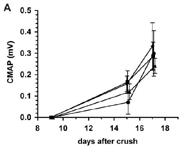
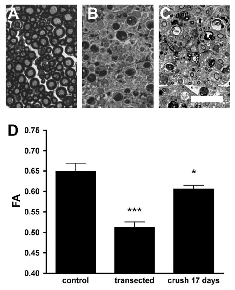
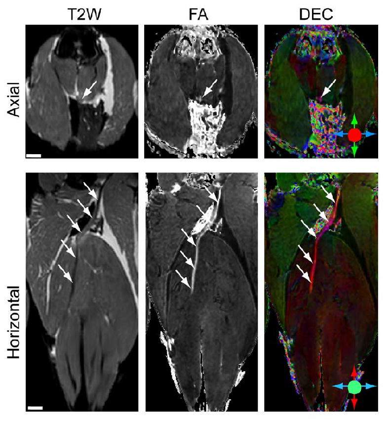
Development of outcome measures to assess ongoing nerve regeneration in the living animal that can be translated to human can provide extremely useful tools for monitoring the effects of therapeutic interventions to promote nerve regeneration. Diffusion tensor imaging (DTI), a magnetic resonance based technique, provides image contrast for nerve tracts and can be applied serially on the same subject with potential to monitor nerve fiber content. In this study, we examined the use of ex vivo high-resolution DTI for imaging intact and regenerating peripheral nerves in mice and correlated the MRI findings with electrophysiology and histology. DTI was done on sciatic nerves with crush, without crush, and after complete transection in different mouse strains. DTI measures, including fractional anisotropy (FA), parallel diffusivity, and perpendicular diffusivity were acquired and compared in segments of uninjured and crushed/transected nerves and correlated with morphometry. A comparison of axon regeneration after sciatic nerve crush showed a comparable pattern of regeneration in different mice strains. FA values were significantly lower in completely denervated nerve segments compared to uninjured sciatic nerve and this signal was restored toward normal in regenerating nerve segments (crushed nerves). Histology data indicate that the FA values and the parallel diffusivity showed a positive correlation with the total number of regenerating axons. These studies suggest that DTI is a sensitive measure of axon regeneration in mouse models and provide basis for further development of imaging technology for application to living animals and humans.
Copyright 2009 Elsevier Inc. All rights reserved.
Figures


Similar articles
-
In vivo evaluation of rabbit sciatic nerve regeneration with diffusion tensor imaging (DTI): correlations with histology and behavior.Magn Reson Imaging. 2015 Jan;33(1):95-101. doi: 10.1016/j.mri.2014.09.005. Epub 2014 Sep 28. Magn Reson Imaging. 2015. PMID: 25271136
-
MRI of peripheral nerve degeneration and regeneration: correlation with electrophysiology and histology.Exp Neurol. 2004 Jul;188(1):171-7. doi: 10.1016/j.expneurol.2004.03.025. Exp Neurol. 2004. PMID: 15191813
-
In vivo evaluation of sciatic nerve crush injury using diffusion tensor imaging: correlation with nerve function and histology.J Comput Assist Tomogr. 2014 Sep-Oct;38(5):790-6. doi: 10.1097/RCT.0000000000000118. J Comput Assist Tomogr. 2014. PMID: 24943253
-
Outcome measures of peripheral nerve regeneration.Ann Anat. 2011 Jul;193(4):321-33. doi: 10.1016/j.aanat.2011.04.008. Epub 2011 May 13. Ann Anat. 2011. PMID: 21640570 Review.
-
Applications of Proteomics to Nerve Regeneration Research.In: Alzate O, editor. Neuroproteomics. Boca Raton (FL): CRC Press/Taylor & Francis; 2010. Chapter 15. In: Alzate O, editor. Neuroproteomics. Boca Raton (FL): CRC Press/Taylor & Francis; 2010. Chapter 15. PMID: 21882439 Free Books & Documents. Review.
Cited by
-
The role of the peripheral and central nervous systems in rotator cuff disease.J Shoulder Elbow Surg. 2015 Aug;24(8):1322-35. doi: 10.1016/j.jse.2015.04.004. J Shoulder Elbow Surg. 2015. PMID: 26189809 Free PMC article. Review.
-
MR Neurography: Advances.Radiol Res Pract. 2013;2013:809568. doi: 10.1155/2013/809568. Epub 2013 Mar 26. Radiol Res Pract. 2013. PMID: 23589774 Free PMC article.
-
Feasibility of simultaneous high-resolution anatomical and quantitative magnetic resonance imaging of sciatic nerves in patients with Charcot-Marie-Tooth type 1A (CMT1A) at 7T.Muscle Nerve. 2022 Aug;66(2):206-211. doi: 10.1002/mus.27647. Epub 2022 Jun 13. Muscle Nerve. 2022. PMID: 35621349 Free PMC article.
-
Three-Dimensional In vivo Magnetic Resonance Imaging (MRI) of Mouse Facial Nerve Regeneration.Front Neurol. 2019 Apr 2;10:310. doi: 10.3389/fneur.2019.00310. eCollection 2019. Front Neurol. 2019. PMID: 31001195 Free PMC article.
-
Declining Skeletal Muscle Function in Diabetic Peripheral Neuropathy.Clin Ther. 2017 Jun;39(6):1085-1103. doi: 10.1016/j.clinthera.2017.05.001. Epub 2017 May 30. Clin Ther. 2017. PMID: 28571613 Free PMC article. Review.
References
-
- Basser P, Mattiello J, LeBihan D. Estimation of the effective self-diffusion tensor from the NMR spin echo. J Magn Reson B. 1994;103:247–254. - PubMed
-
- Basser P, Pajevic S, Pierpaoli C, Duda J, Aldroubi A. In vivo fiber tractography using DT-MRI data. Magn Reson Med. 2000;44:625–632. - PubMed
-
- Basser P, Pierpaoli C. A simplified method to measure the diffusion tensor from seven MR images. Magn Reson Med. 1998;39:928–934. - PubMed
-
- Beaulieu C. The basis of anisotropic water diffusion in the nervous system - a technical review. NMR Biomed. 2002;15:435–455. - PubMed
-
- Beaulieu C, Does M, Snyder R, Allen P. Changes in water diffusion due to Wallerian degeneration in peripheral nerve. Magn Reson Med. 1996;36:627–631. - PubMed
Publication types
MeSH terms
Grants and funding
LinkOut - more resources
Full Text Sources
