. 2010 Jan 12;267(1-3):60-9.
doi: 10.1016/j.tox.2009.10.021. Epub 2009 Oct 29.

The critical role of the cellular thiol homeostasis in cadmium perturbation of the lung extracellular matrix

Affiliations

The critical role of the cellular thiol homeostasis in cadmium perturbation of the lung extracellular matrix

Yinzhi Zhao et al. Toxicology. .

Abstract

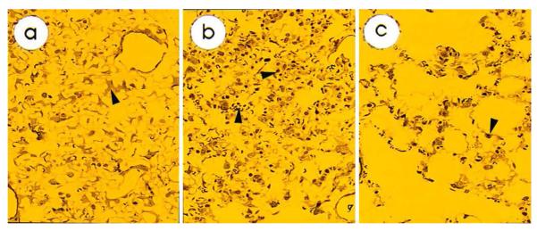
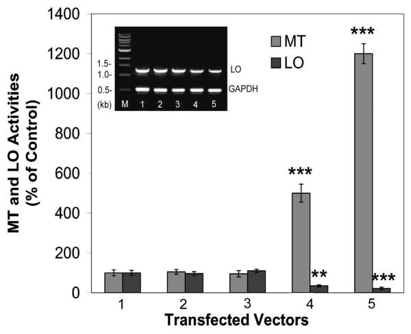
Cadmium (Cd) inhalation can result in emphysema. Cd exposure of rat lung fibroblasts (RFL6) enhanced levels of metal scavenging thiols, e.g., metallothionein (MT) and glutathione (GSH), and the heavy chain of gamma-glutamylcysteine synthetase (gamma-GCS), a key enzyme for GSH biosynthesis, concomitant with downregulation of lysyl oxidase (LO), a copper-dependent enzyme for crosslinking collagen and elastin in the extracellular matrix (ECM). Cd downregulation of LO in treated cells was closely accompanied by suppression of synthesis of collagen, a major structure component of the lung ECM. Using rats intratracheally instilled with cadmium chloride (30 microg, once a week) as an animal model, we further demonstrated that although 2-week Cd instillation induced a non-significant change in the lung LO activity and collagen synthesis, 4- and 6-week Cd instillation resulted in a steady decrease in the lung LO and collagen expression. The lung MT and total GSH levels were both upregulated upon the long-term Cd exposure. Emphysematous lesions were generated in lungs of 6-week Cd-dosed rats. Increases of cellular thiols by transfection of cells with MT-II expression vectors or treatment of cells with GSH monoethyl ester, a GSH delivery system, markedly inhibited LO mRNA levels and catalytic activities in the cell model. Thus, Cd upregulation of cellular thiols may be a critical cellular event facilitating downregulation of LO, a potential mechanism for Cd-induced emphysema.


Figures

Fig. 1
Fig. 1. Cd enhancement of cellular MT and GSH but inhibition of LO activities in cultured RFL6 cells
A. Dose response. Growth-arrested RFL6 cells were exposed to CdCl2 at indicated doses for 24 h. B. Time course. Cells were exposed to 6 μM CdCl2 for various times. The levels of cellular MT, GSH and LO activities in the conditioned media were measured respectively as described in Methods. Data are expressed as % of the corresponding controls (100% MT level in the control = 120 ± 19 ng/mg total cell protein, 100% GSH level in the control = 850 ± 60 ng/mg total cell protein and 100% LO activity in the control = 6,520 ± 580 fluorescence units/mg total cell protein. All values represent the mean ± SD of three experiments each determined with triplicate dishes. *p < 0.05, **p < 0.01, ***p < 0.001 compared with controls without treatment of Cd.
Fig. 2
Fig. 2. Cd stimulation of MT-I, MT-II, and γ-GCS heavy chain expression at protein and mRNA levels
Growth-arrested RFL6 cells were treated with Cd at indicated doses for 24 h, and extracted for preparation of total protein and RNA. Western blot and RT-PCR assays were performed respectively to assess MT-I/MT-II (A and B) and γ-GCS heavy chain (C and D) expression at protein (A and C) and mRNA (B and D) levels. Levels of actin (A and C) and GAPDH (B and D) were determined as internal controls. M in lane 1 of mRNA assays (B and D) represents the DNA molecular marker.
Fig. 3
Fig. 3. Cd inhibition of LO expression at mRNA and protein levels and collagen synthesis
Total protein and RNA were extracted from control and Cd-treated cells, and assayed for the LO mRNA (A) and protein (B) expression by RT-PCR and Western blotting, respectively. For assaying the collagen synthesis, cells were labeled with [14C]proline and incubated in the absence or presence of CdCl2 at indicated doses for 24 h to assess the dose response (C) or at 6 μM CdCl2 for various times to assess the time course response (D). Collagenase-released radioactivities from isolated proteins were assessed by β-counting. In parallel experiment with cells without isotope labeling, LO activities (C and D) in conditioned media were determined using diaminopentane as a substrate and Amplex red as a hydrogen peroxide probe. Data are expressed as % of the control (100% collagen synthesis level in the control = (6.2 ± 0.7) × 104 cpm/106 cells; 100% LO activity in the control = 5,120 ± 515 fluorescence units/mg total cell protein.). All values represent the mean ± SD of three experiments each determined with triplicate dishes. *p < 0.05, **p < 0.01, ***p < 0.001 compared with controls without treatment of Cd.
Fig. 4A-C
Fig. 4A-C. Emphysema-like lesions occurred in repeatedly Cd-exposed rat lungs
Rats (5 rats /group) were exposed to Cd as shown in Methods. Lungs were fixed and processed for routine slice preparation and HE staining. A. HE stained lung tissues under the microscopy at the low magnification (20 x). a and b, the normal lung tissue; c-d, serious inflammatory response in 2-week Cd-instilled lungs as evidenced by occurrence of the leakage of red blood cells in c (arrowhead), the infiltration of inflammatory cells in d (arrowhead); e-h, less inflammatory response in 6-week Cdinstilled lungs coupled with extensive enlargement of airspace (e and f) and multiple pulmonary bullae (g and h) with or without small areas of interstitial fibrosis (arrowheads). B. HE stained lung tissues under the microscopy at the high magnification (100 x). a, the normal lung tissue and the distribution of type II lung epithelial cells (arrowhead); b, serious inflammatory response in 2-week Cd-instilled lungs as evidenced by occurrence of the infiltration of inflammatory cells (arrowhead) and the proliferation of type II lung epithelial cells; c, less inflammatory response found in 6-week Cd-instilled lungs coupled with extensive enlargement of airspace (arrowhead indicates type II lung epithelial cells). C. Electron microscope (EM) examination at the final magnification (3,500 x). a, the normal lung tissues (arrowheads indicate deposition of collagen and elastin in the alveolar wall); b, the lung tissues from 2-week Cd-instilled rats (arrowheads indicate red blood cells and monocyte in the alveolus); c, the lung tissues from 6-week Cd-instilled rats (arrowhead indicates less deposition of collagen and elastin in the alveolar wall). Abbreviations: Alv, alveolus; Cap, capillary; Col, collagen; El, elastin; End C, endothelial cell; Int C, interstitial cell; Mon, monocyte; RBC, red blood cell; Type I LEC, type I lung epithelial cell (arrows); Type II LEC, type II lung epithelial cell; WBC, white blood cell.
Fig. 4D
Fig. 4D. Elevated levels of MT and GSH coupled with decreased LO activity and collagen synthesis in the lung tissue of repeatedly Cd-dosed rats
Rats (5 rats /group) were exposed to Cd as shown in Methods. Physiological saline perfused lung tissues were homogenized and assayed for MT and GSH levels (100% MT level in the control = 381 ± 133 ng/mg lung tissue protein and 100% GSH level in the control = 758 ± 96 ng/mg lung tissue protein). For the LO activity assay, lung tissues were homogenized in 4 M urea and 0.016 M potassium phosphate, pH 7.8. Enzyme activity in lung tissues were assayed using a recombinant human tropoelastin substrate labeled with [3H]lysine (100% LO activity in the control = 2,395 ± 579 cpm of [3H]H2O release/mg lung tissue protein). For the collagen synthesis assay, the physiological saline-perfused lungs were minced into 1 mm3 pieces, and labeled in fresh DMEM supplemented with 100 μCi/ml of [3H]proline for 24 h with gentle shaking. The pulsed tissues were homogenized and centrifuged. Aliquots of supernatants were saved for the protein assay. Pellets washed were resuspended in 0.2 N NaOH and incubated with 100 units/ml ultrapure Clostridial collagenase. The solubilized collagen was separated from noncollagen fraction by 5% TCA precipitation in the presence of 0.25% tannic acid. Radioactivities associated with each fraction (collagen and noncollagen) were determined by β-counting. Collagen synthesis is expressed as collagenase-digestible counts normalized by total protein. Data are expressed as % of the control (100% collagen synthesis level in the control = 6,833 ± 1,377 cpm/mg lung tissue protein). All values represent the mean ± SD (n = or > 3) . *p < 0.05, **p < 0.01, ***p < 0.001 compared with corresponding controls.
Fig. 4E
Fig. 4E. Inhibition of LO expression at protein and mRNA levels in the lung tissue of repeatedly Cd-dosed rats
Physiological saline-perfused lung tissues from control and Cd-dosed rats were extracted with the RIPA buffer and the TRIzol Reagent for preparation of total protein and RNA, respectively. LO protein and mRNA levels in the lung tissues of control (c) and 6-week Cd-instilled rats #1, #2 and #3 (1, 2 and 3) were determined by Western (a) and Northern (b) blots as described. Actin protein and mRNA levels were used as internal controls.
Fig. 5
Fig. 5. Inhibition of LO mRNA expression and catalytic activities by GME, a GSH delivery system, in treated cells
Growth–arrested RFL6 cells were exposed to GME at indicated doses for 24 h followed by washing and incubating in the absence of GME for an additional 24 h period. LO activities in conditioned media and cellular GSH levels were assessed as described in Fig. 1 (100% GSH level in the control = 962 ± 77 ng/mg total cell protein, 100% LO activity in the control = 5,840 ± 771 fluorescence units/mg total cell protein). All values represent the mean ± SD of three experiments each determined with triplicate dishes. *p < 0.05, **p < 0.01, ***p < 0.001 compared with controls without GME treatment. In parallel, total RNA were extracted from control and GME-treated cells, and assayed for the LO mRNA expression by RT-PCR as described in Methods (see the inserted picture).
Fig. 6
Fig. 6. Inhibition of LO mRNA expression and catalytic activities by expression of MT-II cDNA in transfected cells
RFL6 cells were transfected with MT-II expression vectors at indicated doses. Antisense of MT-II expression vectors was used as an internal control. Cells were harvested 48 h thereafter and cellular MT levels and LO activities in the conditioned media were measured as described in Methods. Data were expressed as % of the control in cells cotransfected with pcDNA 3.1 without MT-II cDNA insert (100% MT level in the control = 140 ± 28 ng/mg total cell protein, 100% LO activity in the control = 6,260 ± 814 fluorescence units/mg total cell protein). 1, control cells without transfection; 2, cells tranfected with 6 μg pcDNA3.1 vector without MT-II cDNA insert; 3, cells tranfected with 6 μg pcDNA3.1 vector with MT-II antisense cDNA; 4, cells transfected with 2 μg MT-II cDNA vectors; 5, cells transfected with 6 μg MT-II cDNA vectors. All values represent the mean ± SD of three experiments, each determined with triplicate dishes. *p < 0.05, **p < 0.01, ***p < 0.001 compared with controls. In parallel, total RNA was extracted from control and treated cells, and assayed for the LO mRNA expression by RT-PCR as described in Methods (see the insert picture).

Similar articles

Cited by

References

    1. Anderson ME, Powrie F, Puri RN, Meister A. Glutathione monoethyl ester; preparation, uptake by tissues, and conversion to glutathione. Arch. Biochem. Biophys. 1985;239:538–548. - PubMed
    1. Bedell-Hogan D, Trackman PC, Abrams W, Rosenbloom J, Kagan HM. Oxidation, crosslinking and insolubilization of recombinant tropoelastin by purified lysyl oxidase. J. Biol. Chem. 1993;268:10345–10350. - PubMed
    1. Chen L-J, Zhao Y, Gao S, Chou I-N, Toselli P, Stone P, Li W. Downregulation of lysyl oxidase and upregulation of cellular thiols in rat fetal lung fibroblasts treated with cigarette smoke condensate. Toxicol. Sci. 2005;83:372–379. - PubMed
    1. Chou DK, Zhao Y, Gao S, Chou I-N, Toselli P, Stone P, Li W. Perturbation of copper (Cu) homeostasis and expression of Cu-binding proteins in cadmium-resistant lung fibroblasts. Toxicol Sci. 2007;99:267–276. - PubMed
    1. Davidson JM. Biochemistry and turnover of lung interstitium. Eur. Respir. J. 1990;3:1048–1063. - PubMed

Publication types

MeSH terms