

Review
doi: 10.1016/j.bbapap.2009.10.017.
Epub 2009 Oct 29.
Defining the conserved internal architecture of a protein kinase
Affiliations
- PMID: 19879387
- PMCID: PMC3435107
- DOI: 10.1016/j.bbapap.2009.10.017
Item in Clipboard
Review
Defining the conserved internal architecture of a protein kinase
Biochim Biophys Acta.
2010 Mar.
Abstract
Protein kinases constitute a large protein family of important regulators in all eukaryotic cells. All of the protein kinases have a similar bilobal fold, and their key structural features have been well studied. However, the recent discovery of non-contiguous hydrophobic ensembles inside the protein kinase core shed new light on the internal organization of these molecules. Two hydrophobic "spines" traverse both lobes of the protein kinase molecule, providing a firm but flexible connection between its key elements. The spine model introduces a useful framework for analysis of intramolecular communications, molecular dynamics, and drug design.
Published by Elsevier B.V.
Figures
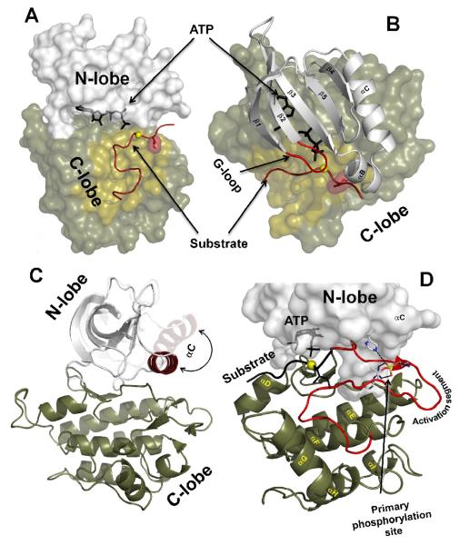
A) Two lobes of a protein kinase molecule (PKA structure 1ATP was used). ATP is bound in a deep cleft between the lobes. The substrate binding groove on the C-lobe is colored sand with the phosphorylation (P) site shown as a yellow sphere. The residues that follow the P-site (P+1 residue) are shown as a dark red surface. B) Five β-strands and a prominent αC-helix from the N-lobe. The G-loop is colored red. C) A large swing motion of the αC-helix is associated with activation (Insulin receptor kinase structures 1IR3 and 1IRK were used). D) Helical content of the C-lobe. The Activation segment is colored red. The primary phosphorylation site, T197 and its binding partners in PKA are shown as sticks.
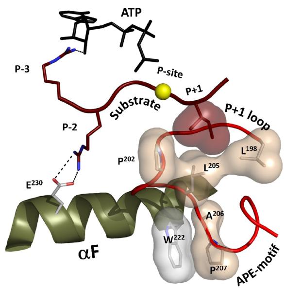
The phosphorylation site for a PKA substrate is shown as a yellow sphere. The hydrophobic pocket created by the P+1 loop is clearly anchored to the conserved W222 in the αF-helix via the APE-motif. The P-2 arginine forms a salt bridge with E230 in the C-terminus of the helix.
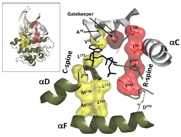
The spines are anchored to the αF-helix and traverse both lobes of the kinase. The C-spine residues are shown as a yellow surface. The R-spine is colored red. The gatekeeper residue is located between the spines (shown as a dark red surface). (Insert: global view of the spines inside the kinase core).
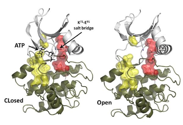
Closed and open conformations of active PKA are shown (PDB IDs 1ATP and 1J3H respectively). The conserved salt bridge between K72 and E91 is formed in the closed configuration and is disrupted in the open state due to the movements in the N-lobe. The spines remain intact during the “breathing” motion of the kinase, which is important for efficient catalysis.
References
-
- Manning G, Plowman GD, Hunter T, Sudarsanam S. Evolution of protein kinase signaling from yeast to man. Trends in Biochemical Sciences. 2002;27:514–520. - PubMed
-
- Knighton DR, Zheng JH, Ten Eyck LF, Ashford VA, Xuong NH, Taylor SS, Sowadski JM. Crystal structure of the catalytic subunit of cyclic adenosine monophosphate-dependent protein kinase. Science. 1991;253:407–414. - PubMed
-
- Adams JA. Activation loop phosphorylation and catalysis in protein kinases: Is there functional evidence for the autoinhibitor model? Biochemistry. 2003;42:601–607. - PubMed
-
- Johnson DA, Akamine P, Radzio-Andzelm E, Madhusudan, Taylor SS. Dynamics of cAMP-Dependent Protein Kinase. Chem. Rev. 2001;101:2243–2270. - PubMed
-
- Johnson LN, Lewis RJ. Structural basis for control by phosphorylation. Chemical Reviews. 2001;101:2209–2242. - PubMed
Publication types
MeSH terms
Substances
Grants and funding
LinkOut - more resources
Full Text Sources
Other Literature Sources
