Review
. 2010 Jan;31(1):7-17.
doi: 10.1016/j.it.2009.09.003. Epub 2009 Oct 31.

T cell-microglial dialogue in Parkinson's disease and amyotrophic lateral sclerosis: are we listening?

Affiliations
Review

T cell-microglial dialogue in Parkinson's disease and amyotrophic lateral sclerosis: are we listening?

Stanley H Appel et al. Trends Immunol. 2010 Jan.

Abstract

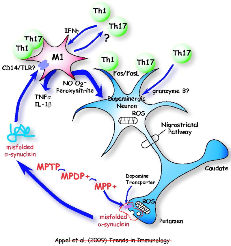
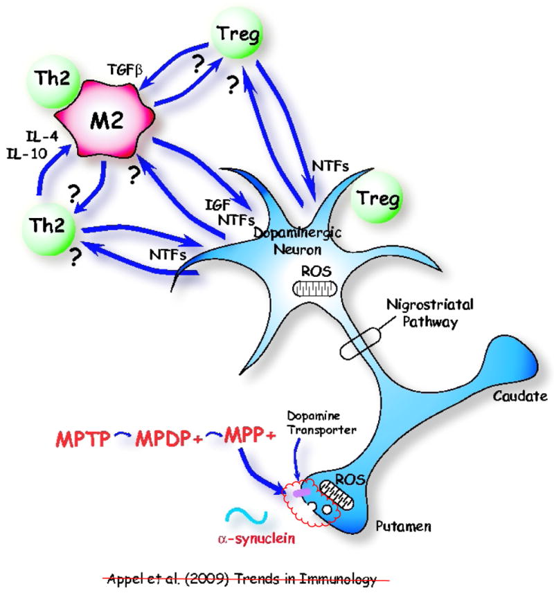
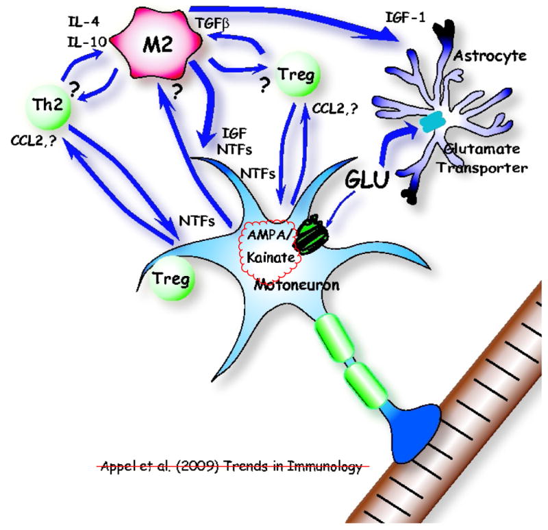
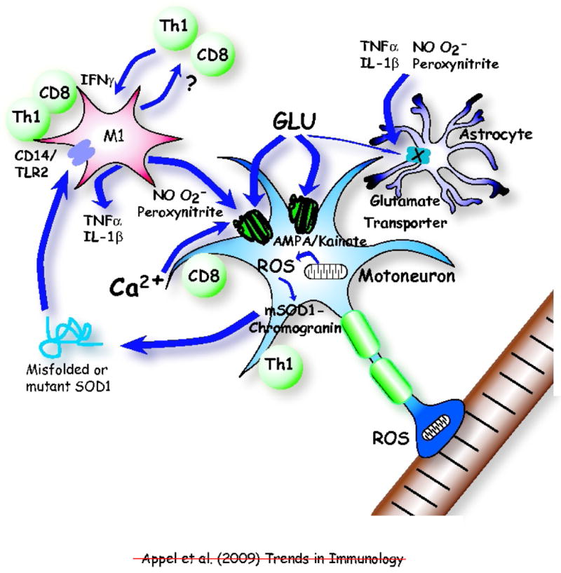
Neuroinflammation is a pathological hallmark in Parkinson's disease (PD) and amyotrophic lateral sclerosis (ALS), and is characterized by activated microglia and infiltrating T cells at sites of neuronal injury. In PD and ALS, neurons do not die alone; neuronal injury is non-cell-autonomous and depends on a well-orchestrated dialogue in which neuronally secreted misfolded proteins activate microglia and initiate a self-propagating cycle of neurotoxicity. Diverse populations and phenotypes of CD4(+) T cells crosstalk with microglia, and depending on their activation status, influence this dialogue and promote neuroprotection or neurotoxicity. A greater understanding of the T cell population that mediates these effects, as well as the molecular signals involved should provide targets for neuroprotective immunomodulation to treat these devastating neurodegenerative diseases.


Figures

Figure 1
Figure 1
Communication between a neuron and the immune system in a chemically induced model of Parkinson's disease. The neurotoxin MPTP is first converted to MPDP+ and then to MPP+ by non-dopaminergic cells; e.g. glia or serotonergic neurons [5]. MPP+ is concentrated in dopaminergic cells via pre-synaptic dopamine transporters (DAT). In the presence of MPP+, reactive oxygen species (ROS) produced by mitochondria in substantia nigra (SN) dopaminergic neurons induce misfolded α-synuclein, which is subsequently released in vesicles at pre-synaptic terminals. The misfolded α-synuclein in turn activates microglia, possibly via a receptor, transforming microglia into a neurotoxic M1 microglial phenotype. Misfolded α-synuclein is released from presynaptic terminals in the striatum and activates microglia in proximity to dopaminergic cell somas in the substantia nigra. It is likely that α-synuclein is also released from dopaminergic cell bodies and activates local microglia, as well as being released from presynaptic dopaminergic terminals and activating adjacent microglia. Potential receptors for misfolded α-synuclein on microglia include CD14/Toll-like receptors (TLRs), and the scavenger receptors. The M1 microglia influence Th1 and perhaps Th17 T cells, which in turn maintain the M1 microglial phenotype via production of IFN-γ. The M1 microglia release ROS nitric oxide (NO) and superoxide radicals (O2•−) form the highly reactive and toxic peroxynitrite. These ROS compound the injury to dopaminergic neurons. M1 microglia also release pro-inflammatory cytokines, such as TNF-α and IL-1β and amplify the toxic inflammation. T cells can also directly injure dopaminergic neurons by signally through the Fas/FasL system.
Figure 2
Figure 2
An immunologically protective model following adaptive transfer of T cells in MPTP-induced Parkinson's disease. Th2 and T regulatory (Treg) cells maintain a neuroprotective M2 microglial phenotype through the release of IL-4, IL-10 and TGF-β. Treg cells suppress microglial synthesis and release of reactive oxygen species (ROS) induced by misfolded α-synuclein. Th2 and Treg cells might also protect neurons directly by cell contact-dependent mechanisms or by releasing brain-derived neurotrophic factor (BDNF) and other neurotrophic factors (NTFs). M2 microglia release insulin-like growth factor-1 (IGF-1) and other NTFs, which protect dopaminergic neurons in the substantia nigra (SN). M2 microglia might also positively influence Th2 and Treg cells. Injured neurons may themselves induce M2 microglia and attract Th2 and Tregs cells via release of chemokines.
Figure 3
Figure 3
The innate and adaptive immune systems help maintain motoneuron homeostasis in an early phase of amyotrophic lateral sclerosis (ALS) as well as in the facial axotomy model. Initially, injured neurons attract and maintain neuroprotective M2 microglia by an unknown signal and attract Th2 and T regulatory (Treg) cells by chemokines, possibly CCL2. The first response to neuronal injury might be neuroprotective, and then with sustained neuronal stress, there is a transformation to a cytotoxic response; the M2 microglial phenotype may well be the CNS initial default state in both Parkinson's disease (PD) and ALS. M2 microglia secrete insulin-like growth factor 1 (IGF-1) and other neurotrophic factors (NTFs), which also enhance the neuroprotective properties of astrocytes; the astrocytic glutamate transporters are functional and actively remove glutamate (GLU). Th2 and Treg cells secrete NTFs, directly protecting motoneurons. IL-4, IL-10, and TGF-β secreted by Th2 and Treg cells help maintain M2 microglia, while M2 microglia also influence Th2 and Treg cells. Treg cells have been shown to directly differentiate macrophages toward the M2 state. M2 microglia may induce suppressive Treg cells (iTreg).
Figure 4
Figure 4
An immunological shift from protection to neurotoxicity accelerates disease in amyotrophic lateral sclerosis (ALS). Initial presentation of mutant superoxide dismutase (mSOD1) or oxidized and misfolded wild-type (WT) SOD1 to T cells might occur in the periphery. Later in disease, as the concentration of extracellular mutant or misfolded SOD1 accumulates, this now rogue protein activates the CD14/Toll-like receptors (TLRs) on microglia transforming them from a protective M2 phenotype to a neurotoxic M1 phenotype. Microglia may also re-present the rogue protein to T cells (Th1 and CD8). In vitro, the M1 microglia release the pro-inflammatory TNF-α and IL-1β cytokines and the reactive oxygen species (ROS) nitric oxide (NO) and superoxide (O2•−), which may form the highly reactive and toxic peroxynitrite. These ROS alter the motoneuron AMPA/kainate channel, resulting in increased glutamate (GLU) susceptibility and subsequent calcium (Ca2+) influx cytotoxicity. Extracellular glutamate concentrations may increase due to reduced numbers and abilities of the glutamate transporters expressed by astrocytes to remove glutamate resulting in further glutamate toxicity. The pro-inflammatory cytokines and ROS may also render the astroglial glutamate transporters ineffective further compounding the Ca2+-induced neurotoxicity. The M1 microglia also influence Th1 and CD8 T cells, which in turn maintain M1 microglial phenotype through the release of IFN-γ and may also exert direct toxic effects on motoneurons.

References

    1. Carson MJ, et al. CNS immune privilege: hiding in plain sight. Immunol Rev. 2006;213:48–65. - PMC - PubMed
    1. Rosen DR, et al. Mutations in Cu/Zn superoxide dismutase gene are associated with familial amyotrophic lateral sclerosis. Nature. 1993;362:59–62. - PubMed
    1. Boillée S, et al. ALS: a disease of motor neurons and their nonneuronal neighbors. Neuron. 2006;52:39–59. - PubMed
    1. Langston JW, et al. Evidence of active nerve cell degeneration in the substantia nigra of humans years after 1-methyl-4-phenyl-1,2,3,6-tetrahydropyridine exposure. Ann Neurol. 1999;46:598–605. - PubMed
    1. Dauer W, Przedborski S. Parkinson's disease: Mechanisms and Models. Neuron. 2003;39:889–909. - PubMed

Publication types