. 2009 Dec 25;284(52):36062-36076.
doi: 10.1074/jbc.M109.064923. Epub 2009 Oct 30.

SHIP2 (SH2 domain-containing inositol phosphatase 2) SH2 domain negatively controls SHIP2 monoubiquitination in response to epidermal growth factor

Affiliations

SHIP2 (SH2 domain-containing inositol phosphatase 2) SH2 domain negatively controls SHIP2 monoubiquitination in response to epidermal growth factor

Julie De Schutter et al. J Biol Chem. .

Abstract

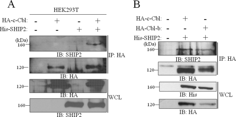
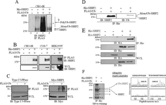
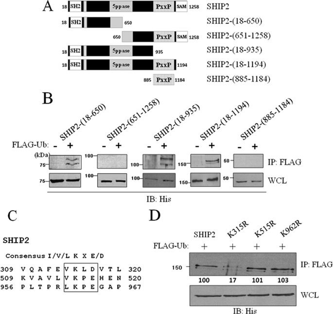
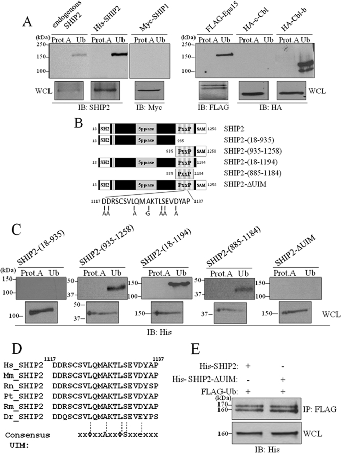
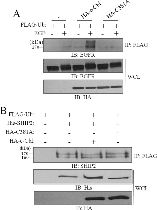
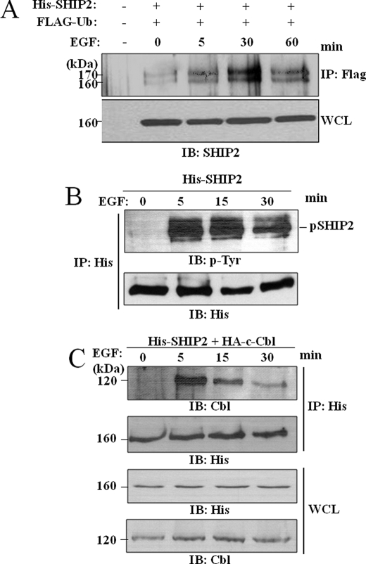
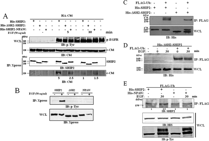
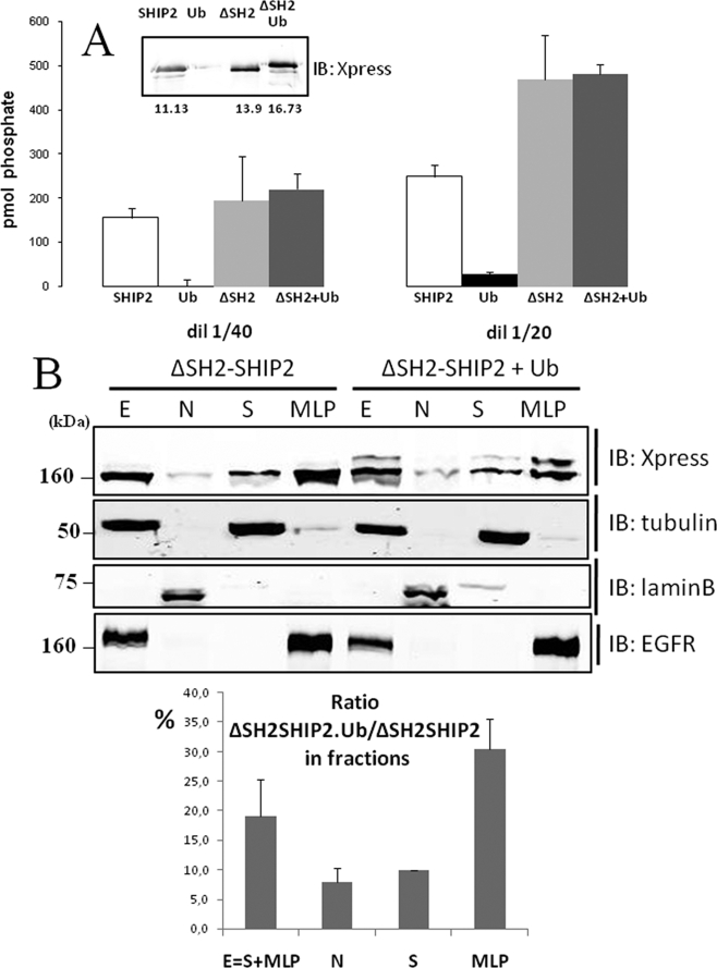
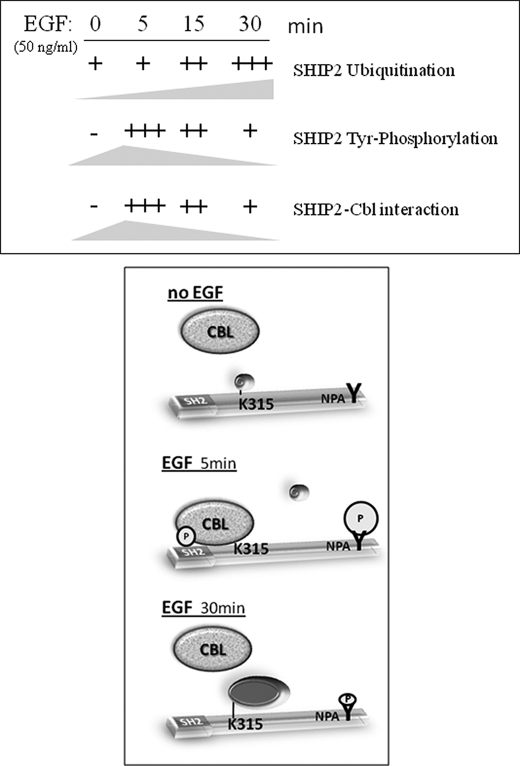
The SH2 domain containing inositol 5-phosphatase SHIP2 contains several interacting domains that are important for scaffolding properties. We and others have previously reported that SHIP2 interacts with the E3 ubiquitin ligase c-Cbl. Here, we identified human SHIP2 monoubiquitination on lysine 315. SHIP2 could also be polyubiquitinated but was not degraded by the 26 S proteasome. Furthermore, we identified a ubiquitin-interacting motif at the C-terminal end of SHIP2 that confers ubiquitin binding capacity. However, this ubiquitin-interacting motif is dispensable for its monoubiquitination. We showed that neither c-Cbl nor Nedd4-1 play the role of ubiquitin ligase for SHIP2. Strikingly, monoubiquitination of the DeltaSH2-SHIP2 mutant (lacking the N-terminal SH2 domain) is strongly increased, suggesting an intrinsic inhibitory effect of the SHIP2 SH2 domain on its monoubiquitination. Moreover, SHIP2 monoubiquitination was increased upon 30 min of epidermal growth factor stimulation. This correlates with the loss of interaction between the SHIP2 SH2 domain and c-Cbl. In this model, c-Cbl could mask the monoubiquitination site and thereby prevent SHIP2 monoubiquitination. The present study thus reveals an unexpected and novel role of SHIP2 SH2 domain in the regulation of its newly identified monoubiquitination.


Figures

FIGURE 1.
FIGURE 1.
SHIP2 associates with c-Cbl and Cbl-b proteins. A, cell lysates were prepared from HEK293T cells transfected with His-tagged SHIP2 and HA-tagged c-Cbl. Appropriate expression of c-Cbl and SHIP2 was confirmed with anti-HA and anti-SHIP2 antibodies, respectively (WCL) IB, immunoblotted; IP, immunoprecipitated. B, cell lysates prepared from COS-7 cells transfected with His-tagged SHIP2 and HA-tagged c-Cbl or HA-tagged Cbl-b were immunoblotted with anti-HA or anti-His antibodies. In both A and B, anti-HA immunoprecipitates were separated by SDS-PAGE and immunoblotted with anti-SHIP2 or anti-HA antibodies.
FIGURE 2.
FIGURE 2.
Monoubiquitination of SHIP2. A, CHO-IR cells were transiently transfected with His-tagged SHIP2 and FLAG-tagged ubiquitin. Anti-FLAG immunoprecipitates were resolved by SDS-PAGE, and endogenous or transfected SHIP2 were both revealed by immunoblotting with anti-SHIP2 antibodies. B, lysates of CHO-IR, COS-7, and HEK293T cells transiently transfected with FLAG-tagged ubiquitin, and His-tagged SHIP2 were immunoprecipitated (IP) with an anti-FLAG antibody and immunoblotted (IB) with anti-SHIP2 antibodies. C, COS-7 cells were transiently transfected with FLAG-tagged ubiquitin and Type I 5-PPase or Myc-tagged SHIP1. Anti-FLAG immunoprecipitates with anti-FLAG antibodies were immunoblotted with anti-Type I 5-PPase or anti-Myc antibodies, respectively. D, CHO-IR cells were transiently transfected with His-tagged SHIP2 and FLAG-tagged ubiquitin. Anti-His immunoprecipitates were resolved by SDS-PAGE and detected by immunoblotting with anti-SHIP2 and anti-ubiquitin (anti-Ub) antibodies. E, COS-7 cells were transiently transfected with HA-tagged SHIP2 and/or His-tagged SHIP2. Anti-His immunoprecipitates were immunoblotted with anti-HA antibodies. F, COS-7 cells were transfected with His-tagged SHIP2 and FLAG-tagged ubiquitin. Anti-His immunoprecipitated proteins were separated by SDS-PAGE and silver stained. Arrows indicate bands of stained proteins excised for enzymatic digestion by trypsin and subsequent analysis by MALDI-Q-TOF mass spectrometry. In-gel tryptic digests of the control SHIP2 (band 1) and the monoubiquitinated SHIP2 (band 2) were analyzed by MALDI-Q-TOF mass spectrometry. Mass spectrometry spectra (enlarged) of the ubiquitin peptides isolated in monoubiquitinated SHIP2 (band 2) are shown. n.s., band is unspecific.
FIGURE 3.
FIGURE 3.
Mapping of the lysine 315 as ubiquitination site in SHIP2. A, deletion mutants of wild-type SHIP2. B, COS-7 cells were transiently transfected with His-tagged SHIP2-(18–650), His-tagged SHIP2-(651–1258), His-tagged SHIP2-(18–935), His-tagged SHIP2-(885–1184), or His-tagged SHIP2-(18–1194) mutants of SHIP2. Cell lysates were immunoprecipitated (IP) with an anti-FLAG antibody and immunoblotted with an anti-His antibody. Expression of SHIP2 mutants was confirmed by immunoblotting of the cell lysates with an anti-His antibody. C, sequence analysis of SHIP2 showed that SHIP2 contains three consensus ubiquitination/sumoylation sites (lysine residues in position 315, 515, and 962). D, COS-7 cells were transiently transfected with FLAG-tagged ubiquitin and His-tagged SHIP2, His-tagged K315R, His-tagged K515R, or His-tagged K962R. Expression of SHIP2 or mutants was confirmed with anti-His antibody. Cell lysates were immunoprecipitated with an anti-FLAG antibody, immunoblotted with an anti-His antibody, and SHIP2 ubiquitination was quantified by densitometric analysis. Each value represents the mean ± S.E. of four independent experiments (*, p < 0.05 compared with control).
FIGURE 4.
FIGURE 4.
SHIP2 binds monoubiquitin by a UIM motif. A, one-fourth lysates of non-transfected COS-7 cells or of COS-7 cells transfected with His-tagged SHIP2, FLAG-tagged Eps15, HA-tagged c-Cbl, HA-tagged Cbl-b, or Myc-tagged SHIP1 were incubated with ubiquitin-agarose (Ub) or protein A-agarose (Prot.A) for 4 h at 4 °C. Bound proteins were analyzed by immunoblotting (IB) with anti-SHIP2, anti-FLAG, anti-HA, or anti-Myc antibodies for the indicated proteins. Expression of transfected proteins was confirmed by immunoblotting of 1/25 lysates with corresponding antibodies. B, schematic representation of the deletion mutants of wild-type SHIP2 we used. C, COS-7 cell lysates transfected with indicated mutants of SHIP2 were incubated with ubiquitin-agarose (Ub) or protein A-agarose (Prot.A) for 4 h at 4 °C. Bound proteins were analyzed by immunoblotting with an anti-His antibody. Expression of transfected proteins was confirmed by immunoblotting of 1/25 lysates with an anti-His antibody. D, alignment of human SHIP2 (amino acids 1117–1137) with its orthologs from different organisms. Consensus UIM is aligned with SHIP2 sequences: Φ, hydrophobic residue; A, alanine; S, serine; and e, acidic residue. E, COS-7 cells were transiently transfected with His-tagged SHIP2 or His-tagged SHIP2-ΔUIM mutant and FLAG-tagged ubiquitin. Cell lysates were immunoprecipitated (IP) with an anti-FLAG antibody and immunoblotted with an anti-His antibody. Expression of transfected SHIP2 proteins was confirmed with an anti-His antibody.
FIGURE 5.
FIGURE 5.
Cbl do not play the role of ubiquitin ligase for SHIP2 monoubiquitination. A, COS-7 cells were transiently transfected with FLAG-tagged ubiquitin and HA-tagged c-Cbl or HA-tagged C381A. Cells were serum-starved for 16 h and stimulated with 50 ng/ml EGF for 10 min. Equal amounts of cell lysates were immunoprecipitated (IP) with an anti-FLAG antibody and immunoblotted (IB) with anti-EGFR antibodies. Expression of EGFR, c-Cbl, and C381A was confirmed with anti-EGFR or anti-HA antibodies. B, COS-7 cells were transiently transfected with His-tagged SHIP2, FLAG-tagged ubiquitin, HA-tagged c-Cbl, HA-tagged C381A, or an empty vector. Cell lysates were immunoprecipitated with an anti-FLAG antibody and immunoblotted with anti-SHIP2 antibodies. Expression of SHIP2, c-Cbl, and c-Cbl-C381A was confirmed with anti-His or anti-HA antibodies.
FIGURE 6.
FIGURE 6.
SHIP2 monoubiquitination is regulated by EGF stimulation. A, COS-7 cells were transiently transfected with FLAG-tagged ubiquitin and His-tagged SHIP2 or an empty vector, serum-starved for 16 h, and stimulated with 50 ng/ml EGF for 5, 30, and 60 min. Equal amounts of cell lysates were immunoprecipitated with an anti-FLAG antibody and immunoblotted with anti-SHIP2 antibodies. Expression of SHIP2 was confirmed by immunoblotting of the cell lysates with anti-SHIP2 antibodies. B and C, COS-7 cells were transfected with His-tagged SHIP2 (B) or with HA-tagged c-Cbl (C), serum-starved for 16 h, and stimulated with 50 ng/ml EGF at 5, 15, and 30 min. Equal amounts of cell lysates were immunoprecipitated (IP) with an anti-His antibody, and immunoprecipitates were separated by SDS-PAGE and immunoblotted (IB) with anti-4G10 (anti-phosphotyrosine) or anti-His antibodies (B) or with anti-Cbl or anti-His antibodies (C).
FIGURE 7.
FIGURE 7.
Loss of the SH2 domain of SHIP2 favors its monoubiquitination. A, COS-7 cells were transiently transfected with HA-Cbl and Xpress-tagged SHIP2, ΔSH2-SHIP2, or NPAW-SHIP2 mutants. Cells are serum-starved for 16 h and stimulated with 50 ng/ml EGF for 5 and 30 min. Cell lysates were immunoprecipitated with an anti-Xpress antibody and immunoblotted (IB) with an anti-HA antibody. The amount of associated c-Cbl was quantified by the Odyssey method. B, COS-7 cells were transiently transfected with Xpress-tagged SHIP2, ΔSH2-SHIP2, or NPAW-SHIP2 mutants. Cells are serum-starved for 16 h and stimulated with 50 ng/ml EGF for 5 min. Phosphorylated SHIP2 was revealed using anti-phosphotyrosine antibody. C, COS-7 cells were transiently transfected with FLAG-tagged ubiquitin, His-tagged SHIP2, or His-tagged ΔSH2-SHIP2 mutant. Cell lysates were immunoprecipitated (IP) with an anti-FLAG antibody and immunoblotted with an anti-His antibody. Expression of His-tagged SHIP2 and ΔSH2-SHIP2 mutant was confirmed with an anti-His antibody. D, COS-7 cells were transiently transfected with FLAG-tagged ubiquitin and His-tagged ΔSH2-SHIP2 mutant as indicated, serum-starved for 16 h, and stimulated with 50 ng/ml EGF for 30 min. Equal amounts of cell lysates were immunoprecipitated with an anti-FLAG antibody and immunoblotted with an anti-His antibody. E, COS-7 cells were transiently transfected with FLAG-tagged ubiquitin and His-tagged SHIP2 or NPAW SHIP2 mutant as indicated, serum-starved for 16 h, and stimulated with 50 ng/ml EGF for 5, 30, and 60 min. Equal amounts of cell lysates were immunoprecipitated with an anti-FLAG antibody and immunoblotted with an anti-SHIP2 antibody. Expression of SHIP2 mutant was confirmed by immunoblotting of the cell lysates with an anti-His antibody, and EGF stimulation was verified using anti-phosphotyrosine antibody.
FIGURE 8.
FIGURE 8.
Catalytic activity and subcellular localization of monoubiquitinated ΔSH2-SHIP2. A, COS-7 cells were transiently transfected with Xpress-tagged ΔSH2-SHIP2 alone or with FLAG-tagged ubiquitin, lysed, and immunoprecipitated overnight with Xpress or FLAG antibodies, respectively. Immunoblotting (IB) with anti-Xpress are shown in the inset. A fraction of the immunoprecipitation (50 and 25 μl) was used to assay phosphatase activity. The results are representative of four different experiments. In each experiments data are means of triplicates ± S.E. B, COS-7 cells were transiently transfected with His-tagged ΔSH2-SHIP2. SHIP2 in each fraction was probed by Western blotting with anti-Xpress antibodies. Tubulin α, lamin B1, and EGFR were used as controls to validate the fractionation. The percentage of ubiquitinated ΔSH2-SHIP2 over non-ubiquitinated SHIP2 in each fraction was estimated by quantitative Western blotting using anti-Xpress antibodies.
FIGURE 9.
FIGURE 9.
Proposed model for the regulation of SHIP2 monoubiquitination. At short times of EGF stimulation (5 min), SHIP2 would be protected from monoubiquitination due to the integrity of its SH2 domain, which could be engaged in an intramolecular interaction with an internal phosphorylated tyrosine or through interaction with a phosphorylated tyrosine of a proteic partner. After longer EGF stimulation times (30 min), disruption of the intramolecular interaction, caused by decreasing of SHIP2 tyrosine phosphorylation, or disruption of interaction with a partner would allow the accessibility of the monoubiquitination site and favor the SHIP2 monoubiquitination. The labels used are as follows: SH2, SHIP2 SH2 domain; K315, lysine on position 315 in SHIP2; and pY, phosphorylated tyrosine.

Similar articles

Cited by

References

    1. Ooms L. M., Horan K. A., Rahman P., Seaton G., Gurung R., Kethesparan D. S., Mitchell C. A. (2009) Biochem. J. 419, 29–49 - PubMed
    1. McCrea H. J., De Camilli P. (2009) Physiology 24, 8–16 - PMC - PubMed
    1. Pesesse X., Deleu S., De Smedt F., Drayer L., Erneux C. (1997) Biochem. Biophys. Res. Commun. 239, 697–700 - PubMed
    1. Ishihara H., Sasaoka T., Hori H., Wada T., Hirai H., Haruta T., Langlois W. J., Kobayashi M. (1999) Biochem. Biophys. Res. Commun. 260, 265–272 - PubMed
    1. Pesesse X., Dewaste V., De Smedt F., Laffargue M., Giuriato S., Moreau C., Payrastre B., Erneux C. (2001) J. Biol. Chem. 276, 28348–28355 - PubMed

Publication types

MeSH terms

Substances

LinkOut - more resources