. 2010 Feb;33(2):350-5.
doi: 10.2337/dc09-1141. Epub 2009 Oct 30.

Proinflammatory modulation of the surface and cytokine phenotype of monocytes in patients with acute Charcot foot

Affiliations

Proinflammatory modulation of the surface and cytokine phenotype of monocytes in patients with acute Charcot foot

Luigi Uccioli et al. Diabetes Care. 2010 Feb.

Abstract

Objective: Despite increased information on the importance of an inappropriate inflammatory response in the acute Charcot process, there has been no previous attempt to define the specific pathways that mediate its pathogenesis. Here, the role played by monocytes was analyzed.

Research design and methods: The immune phenotype of peripheral monocytes was studied by fluorescence-activated cell sorter analysis comparing patients with acute Charcot (n = 10) in both the active and recovered phase, diabetic patients with neuropathy (with or without osteomyelitis), and normal control subjects.

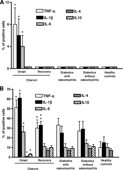
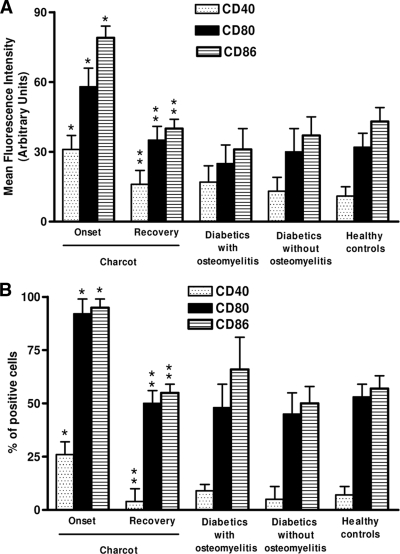
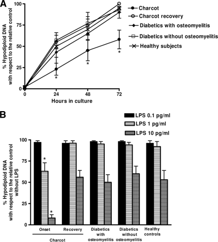
Results: When compared with diabetic control subjects and healthy subjects, monocytes from acute Charcot patients showed a proinflammatory immune phenotype characterized by increased production of proinflammatory cytokines, reduced secretion of anti-inflammatory cytokines, increased expression of surface costimulatory molecules, and increased resistance to serum withdrawal-induced apoptosis. In addition, the pattern of circulating cytokines confirmed activation of proinflammatory cytokines. No modulation of the monocyte phenotype was documented in diabetic control subjects and healthy subjects, thus indicating that the proinflammatory alterations of monocytes are specific and causative of acute Charcot.

Conclusions: Together, these data provide evidence for the role of proinflammatory changes in the immune phenotype of monocytes in the pathogenesis of acute Charcot. These alterations may explain the abnormally intense and prolonged inflammatory response that characterizes this disorder and may represent a potential therapeutic target for specific pharmacological interventions.


Figures

Figure 1
Figure 1
Spontaneous cytokine production by monocytes from acute Charcot case subjects and control subjects. Spontaneous (A) and inducible (monocytes stimulated for 18 h by 100 ng/ml LPS) (B) cytokine production was assessed by FACS, as intracellular accumulation on a single-cell basis. Appropriate controls with isotype-matched irrelevant mAbs were carried out and consistently showed <1% of positive cells. For each analysis, 104 monocytes were gated according to scatter characteristics designed to include only viable cells. Fluorescence data were expressed as percentage of positive cells after subtraction of background isotype-matched values. The data represent the means ± SD (error bars). *P < 0.05, with respect to control subjects. **P > 0.005, with respect to diabetic control and healthy subjects.
Figure 2
Figure 2
Upmodulation of surface molecules in monocyte-macrophages from Charcot patients. The expression of CD40, CD80, and CD86 was analyzed by FACS on CD14+ gated cells in fresh explanted PBMCs. Appropriate controls with isotype-matched irrelevant mAbs were carried out and consistently showed <1% of positive cells. For each analysis, 104 monocytes were gated according to scatter characteristics designed to include only viable cells. Fluorescence data were expressed as mean channel fluorescence (A) and as percentage of positive cells (B) after subtraction of background isotype-matched values. The data represent the means ± SD (error bars). *P < 0.05, with respect to control subjects; **P > 0.05, with respect to diabetic control and healthy subjects.
Figure 3
Figure 3
Kinetics of hypodiploid DNA formation in monocyte-macrophages. PBMCs were cultured in medium with no added serum and hypodiploid DNA formation was determined at indicated intervals (A). PBMCs were cultured in medium with no added serum with different LPS concentrations, and hypodiploid DNA formation was determined at 72 h (B). The red fluorescence due to propidium iodide staining of the DNA was registered on a logarithmic scale at >620 nm. The forward and side scatter of particles were simultaneously measured. Cell debris was excluded from analysis by appropriately raising the forward scatter threshold. The residual cell debris had a very low DNA fluorescence emission and a low side scatter signal. At least 104 cells of each sample were analyzed. *P < 0.05, with respect to recovered acute case subjects and control subjects.

References

    1. La Fontaine J, Harkless LB, Sylvia VL, Carnes D, Heim-Hall J, Jude E. Levels of endothelial nitric oxide synthase and calcitonin gene-related peptide in the Charcot foot: a pilot study. J Foot Ankle Surg 2008; 47: 424– 429 - PubMed
    1. Mabilleau G, Petrova NL, Edmonds ME, Sabokbar A. Increased osteoclastic activity in acute Charcot's osteoarthropathy: the role of receptor activator of nuclear factor-κβ ligand. Diabetologia 2008; 51: 1035– 1040 - PMC - PubMed
    1. Jeffcoate W. Vascular calcification and osteolysis in diabetic neuropathy: is RANKL the missing link? Diabetologia 2004; 47: 1488– 1492 - PubMed
    1. Jeffcoate WJ, Game F, Cavanagh PR. The role of proinflammatory cytokines in the cause of neuropathic osteoarthropathy (acute Charcot foot) in diabetes. Lancet 2005; 366: 2058– 2061 - PubMed
    1. Lam J, Abu-Amer Y, Nelson CA, Fremont DH, Ross FP, Teitelbaum SL. Tumor necrosis factor superfamily cytokines and the pathogenesis of inflammatory osteolisis. Ann Rheum Dis 2002; 62( Suppl. 2): ii82– ii83 - PMC - PubMed

MeSH terms