. 2010 Jan;192(1):155-68.
doi: 10.1128/JB.01260-09.

A type VI secretion system effector protein, VgrG1, from Aeromonas hydrophila that induces host cell toxicity by ADP ribosylation of actin

Affiliations

A type VI secretion system effector protein, VgrG1, from Aeromonas hydrophila that induces host cell toxicity by ADP ribosylation of actin

G Suarez et al. J Bacteriol. 2010 Jan.

Abstract

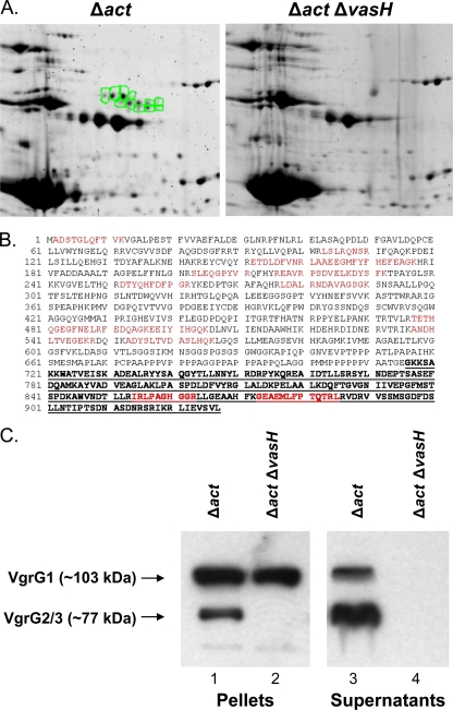
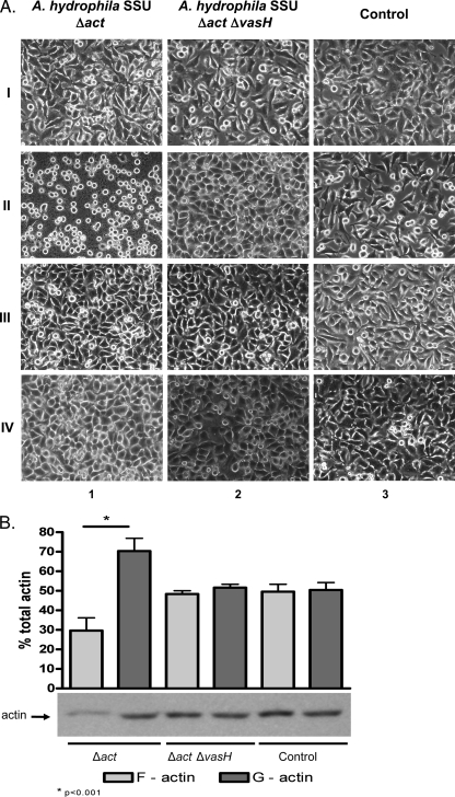
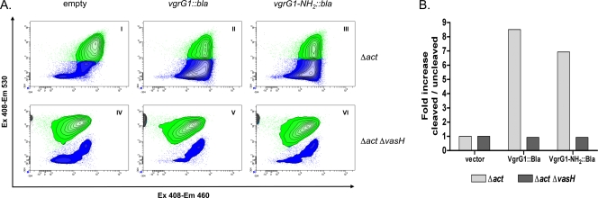
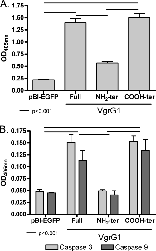
We recently delineated the importance of a type VI secretion system (T6SS) gene cluster in the virulence of diarrheal isolate SSU of Aeromonas hydrophila and showed that VasH, a sigma(54) activator and T6SS component, was involved in the production of its associated effectors, e.g., hemolysin-coregulated protein. To identify additional T6SS effectors and/or secreted proteins, we subjected culture supernatants from deletion mutants of A. hydrophila, namely, a Delta act mutant (a T2SS-associated cytotoxic enterotoxin-encoding gene) and a Delta act Delta vasH mutant, to 2-dimensional gel electrophoresis and mass spectrometric analysis. Based on these approaches, we identified a member of the VgrG protein family, VgrG1, that contained a vegetative insecticidal protein (VIP-2) domain at its carboxyl-terminal end. Consequently, the vgrG1 gene was cloned in pBI-EGFP and pET-30a vectors to be expressed in HeLa Tet-Off cells and Escherichia coli, respectively. We assessed the ADP-ribosyltransferase (ADPRT) activity of various domains of purified recombinant VgrG1 (rVgrG1) and provided evidence that only the full-length VgrG1, as well as its carboxyl-terminal domain encoding the VIP-2 domain, showed ADPRT activity. Importantly, bacterium-host cell interaction was needed for the T6SS to induce cytotoxicity in eukaryotic cells, and we demonstrated translocation of VgrG1. Furthermore, our data indicated that expression of the genes encoding the full-length VgrG1 and its carboxyl-terminal domain in HeLa Tet-Off cells disrupted the actin cytoskeleton, which was followed by a decrease in cell viability and an increase in apoptosis. Taken together, these findings demonstrated for the first time that VgrG1 of A. hydrophila possessed actin ADPRT activity associated with its VIP-2 domain and that this domain alone was able to induce a rounded phenotype in HeLa Tet-Off cells, followed by apoptosis mediated by caspase 9 activation.


Figures

FIG. 1.
FIG. 1.
Identification of proteins secreted via T6SS in the supernatant of A. hydrophila SSU. (A) Comparison of 2-D gels containing proteins from the supernatants of the A. hydrophila SSU Δact (left) and Δact ΔvasH (right) mutant strains. Highlighted spots (in green) represent a cluster of proteins secreted via the T6SS which were identified by mass spectrometric analysis. (B) Alignment of VgrG1 from A. hydrophila ATCC 7966 (gi|117619461) with peptides identified via mass spectrometry (red) in one of the secreted proteins (VgrG1) from A. hydrophila SSU. The bold, underlined sequence represents the VIP-2 domain. (C) Western blot analysis of pellet and supernatant fractions of A. hydrophila SSU Δact (lanes 1 and 3) and Δact ΔvasH (lanes 2 and 4) mutant strains using antibodies specific to the VIP-2 domain of VgrG1 in combination with specific antibodies against VgrG2.
FIG. 2.
FIG. 2.
VgrG1 has ADP-ribosyltransferase (ADPRT) activity, which is associated with the presence of the VIP-2 domain. ADPRT assays were performed by using 6-biotin-17-NAD, purified rVgrG proteins, and HeLa cell lysates (A) or recombinant nonmuscle actin (B) as a source of target protein. The reaction mixtures were separated on SDS-12% PAGE gels and electroblotted to nitrocellulose membranes, and the incorporation of biotin-ADP by the target protein was detected by using streptavidin-HRP. The nature of the sample loaded in each lane of the gels is depicted in a table below panel B.
FIG. 3.
FIG. 3.
(A) Induction of HeLa rounded-cell phenotype in cocultures with different strains of A. hydrophila SSU. HeLa cells were cocultured for 90 min with the A. hydrophila SSU Δact mutant (column 1) and Δact ΔvasH mutant (column 2) or cultured alone (noninfected HeLa cell control; column 3) in direct bacterium-host cell contact (II) or by using transwell inserts (III). Supernatants from cocultures in direct cell-to-cell contact were collected after 90 min and used as preconditioned media on fresh HeLa cell cultures (IV). The initial morphology of HeLa cells at 0 min is shown in row I, and that of noninfected HeLa cells (control) in column 3. Original magnification, ×400. (B) Quantification of G- and F-actin by Western blot and densitometric analyses. HeLa cells in direct contact with different strains of A. hydrophila SSU were harvested after 90 min of coculture. Cells were lysed and processed as indicated in Materials and Methods. The bar graph represents percentages (means ± standard deviations) of G- and F-actin in HeLa cells infected with different mutant strains from three independent experiments, and the Western blot image is representative of all of them.
FIG. 4.
FIG. 4.
Translocation of VgrG1 into HeLa cell cytoplasm. HeLa cells were infected with the A. hydrophila SSU Δact or Δact ΔvasH mutant strain expressing and producing full-length VgrG1::Bla (referred to as VgrG1::Bla) or VgrG1-NH2::Bla. As a control, HeLa cells were infected with bacteria containing the empty vector. (A) Flow cytometric density plots showing disruption of CCF4 FRET (from green to blue) due to translocation of Bla into cytoplasm of HeLa cells infected with the A. hydrophila Δact parental strain (II and III) compared to its disruption in host cells infected with A. hydrophila SSU containing the empty vector (I and IV). HeLa cells infected with the A. hydrophila Δact ΔvasH mutant expressing and producing the fusion proteins were not able to translocate Bla and did not disrupt the CCF4 FRET (V and VI). For analysis, 2 × 10⁵ HeLa cells were acquired and gated in side forward/side scatter patterns to avoid aggregates. Ex, excitation wavelength; Em, emission wavelength. (B) Fold increases in percentages of blue HeLa cells with cleaved substrate versus green HeLa cells with uncleaved substrate (after infection with A. hydrophila SSU Δact and Δact ΔvasH mutant strains) compared to the results for HeLa cells infected with bacteria carrying the empty vector. The graph shows data from a representative experiment.
FIG. 5.
FIG. 5.
(A) Western blot analysis of HeLa Tet-Off cell lysates expressing and producing different fragments of VgrG1. The various forms of VgrG1 were detected by using two types of sera. We used sera from mice immunized with rVgrG2 of A. hydrophila SSU which cross-reacted with the NH2-terminal portion of VgrG1 (left panel) and from mice immunized with the recombinant VIP-2 domain of VgrG1 of A. hydrophila ATCC 7966 which recognized only VgrG1 and its COOH-terminal domain. The samples loaded in the lanes are depicted on the top. α, anti. (B) Morphological changes of HeLa Tet-Off cells induced by the expression of different VgrG1 fragments of A. hydrophila ATCC 7966. Host cells were stained for actin cytoskeleton by using Alexa Fluor 568-phalloidin (red), and the expression of the enhanced fluorescent green protein (EGFP) gene was detected in cells successfully transfected with the pBI-EGFP vector alone (I) or with the vector containing genes encoding NH2-terminal (II), full-length (III), or COOH-terminal (IV) fragments of VgrG1. The blue strain (DAPI) shows nuclei. Original magnification, ×400. (C) Quantification of actin cytoskeleton (F-actin) as measured by fluorescent phalloidin staining of HeLa Tet-Off cells expressing and producing different VgrG1 fragments. Flow cytometry dot plots show results for HeLa Tet-Off cells stained with Alexa Fluor 568-phalloidin and expressing different fragments of VgrG1. The analysis was performed on EGFP-positive cells. The percentages of positive cells from representative experiments (72 h) are shown in the plotted quadrants (left), and mean fluorescent intensity values (MFI) from three different assays at 24 h and 72 h were determined (right). Statistical differences at 24 h (P < 0.01) and 72 h (P < 0.001) were noted between results for cells expressing vector alone (pBI-EGFP) and cells expressing and producing full-length VgrG1 and the COOH-terminal fragment and between results for cells expressing and producing the NH2-terminal fragment and cells expressing full-length VgrG1 and the COOH-terminal fragment. (D) Quantification of F-actin and G-actin present in HeLa Tet-Off cells expressing and producing different VgrG1 fragments. The percentages of F- and G-actin per sample (30 μl) were analyzed by Western blot analysis and by using antibodies to actin followed by densitometric scanning of the blots. Densitometric quantification of the results from three different assays and a representative Western blot image are shown. Statistical differences at 24 h (P < 0.001) were noted between fractions containing F- and G-actin in HeLa cells expressing full-length VgrG1 and the COOH-terminal fragments. Asterisks indicate statistically significant differences. The designation ter refers to NH2- or COOH-terminal domains.
FIG. 6.
FIG. 6.
Viability of HeLa Tet-Off cells expressing genes encoding different fragments of VgrG1 from A. hydrophila ATCC 7966. Percentages of dead and/or dying cells were quantified by incorporation of 7-AAD and flow cytometry of HeLa Tet-Off cells expressing vector alone (pBI-EGFP) or producing different fragments of VgrG1 (NH2- and COOH-terminal domains and full-length) after 24 h and 72 h of transfection. Means ± standard deviations of the results from three different assays are shown, and statistical significance is indicated. The designation ter refers to NH2- or COOH-terminal domains.
FIG. 7.
FIG. 7.
Apoptosis of HeLa Tet-Off cells expressing genes encoding different fragments of VgrG1. Apoptosis rates were measured by quantification of cytoplasmic nucleosomes (A) and levels of caspase 3 and caspase 9 activity (B) in lysates of HeLa Tet-Off cells expressing vector alone (pBI-EGFP) or genes encoding NH2- and COOH-terminal fragments and full-length VgrG1 from A. hydrophila ATCC 7966. Means ± standard deviations of the results from three different assays are shown, and statistical significance (P < 0.001) is indicated. The designation ter refers to NH2- or COOH-terminal domains. OD405nm, optical density at 405 nm.

References

    1. Aktories, K., M. Barmann, I. Ohishi, S. Tsuyama, K. H. Jakobs, and E. Habermann. 1986. Botulinum C2 toxin ADP-ribosylates actin. Nature 322:390-392. - PubMed
    1. Aktories, K., and A. Wegner. 1989. ADP-ribosylation of actin by clostridial toxins. J. Cell Biol. 109:1385-1387. - PMC - PubMed
    1. Aktories, K., U. Weller, and G. S. Chhatwal. 1987. Clostridium botulinum type C produces a novel ADP-ribosyltransferase distinct from botulinum C2 toxin. FEBS Lett. 212:109-113. - PubMed
    1. Albert, M. J., M. Ansaruzzaman, K. A. Talukder, A. K. Chopra, I. Kuhn, M. Rahman, A. S. Faruque, M. S. Islam, R. B. Sack, and R. Mollby. 2000. Prevalence of enterotoxin genes in Aeromonas spp. isolated from children with diarrhea, healthy controls, and the environment. J. Clin. Microbiol. 38:3785-3790. - PMC - PubMed
    1. Barth, H., K. Aktories, M. R. Popoff, and B. G. Stiles. 2004. Binary bacterial toxins: biochemistry, biology, and applications of common Clostridium and Bacillus proteins. Microbiol. Mol. Biol. Rev. 68:373-402. - PMC - PubMed

Publication types

MeSH terms