Review
. 2010 Mar;139(3):479-93.
doi: 10.1530/REP-09-0255. Epub 2009 Oct 30.

Spermatogonial stem cells in higher primates: are there differences from those in rodents?

Affiliations
Review

Spermatogonial stem cells in higher primates: are there differences from those in rodents?

Brian P Hermann et al. Reproduction. 2010 Mar.

Abstract

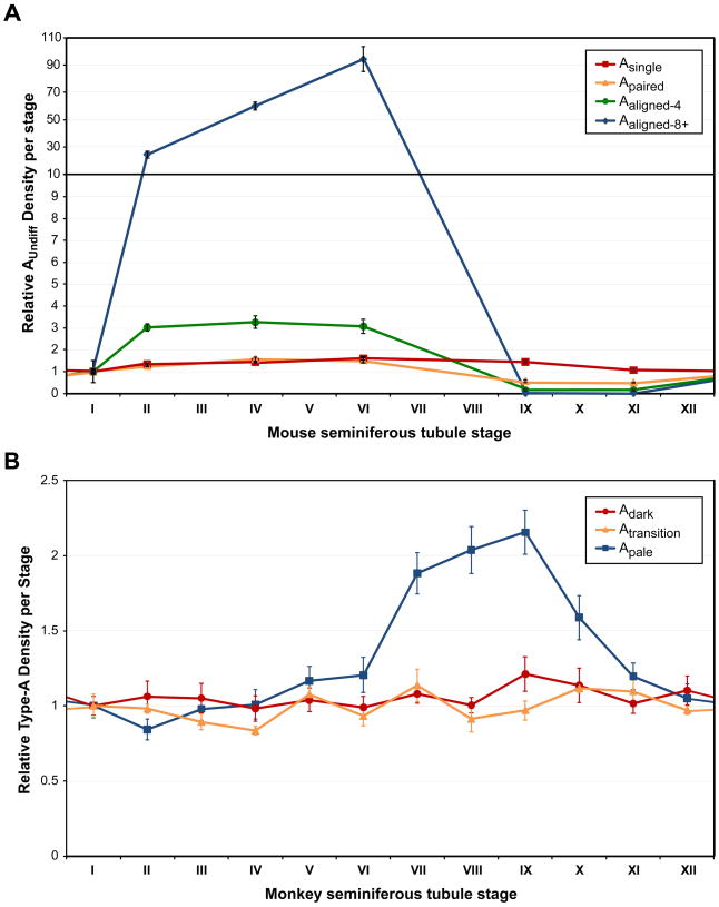
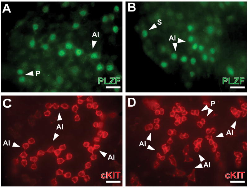
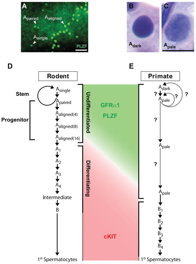
Spermatogonial stem cells (SSCs) maintain spermatogenesis throughout the reproductive life of mammals. While A(single) spermatogonia comprise the rodent SSC pool, the identity of the stem cell pool in the primate spermatogenic lineage is not well established. The prevailing model is that primate spermatogenesis arises from A(dark) and A(pale) spermatogonia, which are considered to represent reserve and active stem cells respectively. However, there is limited information about how the A(dark) and A(pale) descriptions of nuclear morphology correlate with the clonal (A(single), A(paired), and A(aligned)), molecular (e.g. GFRalpha1 (GFRA1) and PLZF), and functional (SSC transplantation) descriptions of rodent SSCs. Thus, there is a need to investigate primate SSCs using criteria, tools, and approaches that have been used to investigate rodent SSCs over the past two decades. SSCs have potential clinical application for treating some cases of male infertility, providing impetus for characterizing and learning to manipulate these adult tissue stem cells in primates (nonhuman and human). This review recounts the development of a xenotransplant assay for functional identification of primate SSCs and progress dissecting the molecular and clonal characteristics of the primate spermatogenic lineage. These observations highlight the similarities and potential differences between rodents and primates regarding the SSC pool and the kinetics of spermatogonial self-renewal and clonal expansion. With new tools and reagents for studying primate spermatogonia, the field is poised to develop and test new hypotheses about the biology and regenerative capacity of primate SSCs.


Conflict of interest statement

Declaration of Interest: The authors have no conflicts of interest to declare.

Figures

Figure 1
Figure 1. Relative distribution of undifferentiated Type-A spermatogonia in mice and monkeys
The relative number of undifferentiated spermatogonia in any given segment of seminiferous tubules at each stage of the seminiferous cycle are shown for (A) mice (Tegelenbosch & de Rooij 1993) and (B) monkeys (Fouquet & Dadoune 1986). For A, data for the relative number of Asingle (Red squares, line), Apaired (Orange triangles, line), and Aaligned clones of 4 and 8+ cells (green circles, blue diamonds) are shown for C3H-101 F1 hybrid mice (Tegelenbosch & de Rooij 1993). The calculated relative density of undifferentiated Type-A spermatogonia at each stage was determined by dividing the value for each stage (number of spermatogonia per 1000 Sertoli cells) by the same value for Stage I (Tegelenbosch & de Rooij 1993). In B, data for the relative density of Adark, Atransition and Apale per stage is as reported for cynomolgus monkeys (Fouquet & Dadoune 1986). The calculated relative density of Type-A spermatogonia in each stage (number of spermatogonia per 100 Sertoli cells) was normalized to the value for Stage I (Fouquet & Dadoune 1986).
Figure 2
Figure 2. SSC transplantation from different donor species into busulfan-treated mouse testes
(A–B) Donor GFP mouse testis cells (green) at 2 weeks after transplantation. Margins of recipient seminiferous tubules are marked by a dashed white line. (CD) Donor GFP mouse testis cells (green) at 2 months after transplantation. Patches of transplanted donor (EF) rhesus and (GH) human testis cells in immune-deficient nude mouse seminiferous tubules were detected by whole-mount immunohistochemistry using the rhesus testis cell antibody. Scale bars = 50μm. Adapted from (Hermann et al. 2007; Hermann et al. 2009) and unpublished data.
Figure 3
Figure 3. Correlation between molecular markers of rodent SSCs (GFRα1, PLZF, NGN3, cKIT) and morphological descriptions (Adark, Apale, B) of spermatogonia in the adult rhesus testis
Sections of adult rhesus testes were evaluated by immunohistochemistry for GFRα1, PLZF, NGN3, and cKIT (Hermann et al. 2009). Subsequently, sections were counterstained by the PAS-Hematoxylin method to reveal nuclear morphology and identify Adark and Apale spermatogonia, as well as differentiating Type-B spermatogonia. (A–D) Representative staining for GFRα1 is shown. The image in (A) shows part of one seminiferous tubule (scale bar = 50μm). Enlargements are also shown of representative Adark (B), Apale (C) and B4 spermatogonia (D) (scale bar = 10μm). For all spermatogonia that could be definitively classified as Adark or Apale, the percentage that were labeled for GFRα1 is shown. (E) Colored bars (GFRα1, blue; PLZF, green; NGN3, black; cKIT, red) indicate the extent of marker expression in the adult spermatogenic lineage based on recently published data (Hermann et al. 2009) (rhesus, Top) or previously published mouse studies [reviewed in (Hermann et al. 2009)] (mice, Bottom). Colored boxes indicate functional descriptors “Stem” (orange), “Progenitor” (yellow) and “Differentiating” (violet), based on rodent data and may identify rhesus spermatogonia with corresponding phenotype and function. The transitions from stem to progenitor or progenitor to differentiating are noted by gradient shading in the middle grey bar between these functional categories. The following abbreviations are used for rodents: As = Asingle, Apr = Apaired, Aal = Aaligned. Adapted from (Hermann et al. 2009).
Figure 4
Figure 4. Comparative immunohistochemical analysis reveals species-specific staining profiles for the stem/progenitor marker PLZF
To begin translating knowledge of rodent and monkey SSCs to humans, we have initiated comparative marker analysis using immunohistochemistry for the transcription factor PLZF in sections from (AB) mouse, (DE) rhesus macaque, and (GH) human testes. Images of sections incubated with non-immune isotype control IgGs are also shown for (C) mouse, (F) monkey, and (I) human to demonstrate non-specific background staining and tissue autofluorescence. White asterisks in A & C indicate the non-specific fluorescent signal observed in interstitial space between seminiferous tubules. Images are shown from (A, C, D, F, G, I) low magnification and (B, D, F) high magnification. PLZF immunoreactivity was observed as a nuclear fluorescent signal (green) in all three species. Sections were counterstained with DAPI (blue). Scale bar = 50μm. (Hermann, Hansel and Orwig, unpublished).
Figure 5
Figure 5. Clonal organization of undifferentiated and differentiating spermatogonia in adult rhesus seminiferous tubules
Determining the clonal arrangement of undifferentiated and differentiating spermatogonia may be possible using whole mount immunohistochemistry in intact seminiferous tubules. In separate experiments, (AB) undifferentiated (PLZF+, Green) or (CD) differentiating (cKIT+, Red) spermatogonia were detected in adult rhesus seminiferous tubules. Some clones are identified in each panel as single (S), pairs (P) or aligned (Al). Scale bars = 25μm. (Hermann and Orwig, unpublished). Note: this is not a co-staining experiment.
Figure 6
Figure 6. Current conceptual models of rodent and primate spermatogenesis
(A) Immunohistochemical staining for PLZF (green) was performed using whole-mount preparations of adult rat seminiferous tubules and clones of PLZF+ spermatogonia were identified as Asingle, Apaired, or Aaligned using the 25μm criteria (Huckins 1971; de Rooij & Russell 2000). Scale bar = 50μm. Sections of rhesus macaque testes were stained using the periodic acid-Schiff method and counterstained with Gills hematoxylin (Simorangkir et al. 2003) to reveal nuclear morphology and identify (B) Adark and (C) Apale spermatogonia. Scale bars = 10μm. (D) Rodent undifferentiated spermatogonia are noted (bracket) including stem spermatogonia (SSCs) comprised of Asingles and some Apaired spermatogonia that will complete cytokinesis to produce new Asingles and maintain the stem cell pool. Transit-amplifying progenitors include some Apaired and Aaligned spermatogonia (chains of 4–16 cells). Whole mount and transplantation analyses provided phenotypes for cells in these categories: stem (Asingle and some Apaired; GFRα1+, PLZF+, and cKIT), progenitor (some Apaired and Aaligned; GFRα1+, PLZF+, cKIT+/−) and differentiating (A1–4, Intermediate, B; GFRα1, PLZF, and cKIT+). (E) In primate testes, undifferentiated (Type-A) spermatogonia are designated Adark and Apale based on nuclear architecture and staining intensity with hematoxylin. Recent progress has provided information about the molecular phenotype of Adark and Apale spermatogonia that allow alignment with rodent spermatogonia exhibiting a similar phenotype. The number of Apale generations is still not clear and may be resolved by future whole-mount marker and clone size analysis (dotted arrows with question marks).

References

    1. Amann RP. The cycle of the seminiferous epithelium in humans: a need to revisit? J Androl. 2008;29:469–487. - PubMed
    1. Barroca V, Lassalle B, Coureuil M, Louis JP, Le PF, Testart J, Allemand I, Riou L, Fouchet P. Mouse differentiating spermatogonia can generate germinal stem cells in vivo. Nat Cell Biol. 2009;11:190–196. - PubMed
    1. Bartmanska J, Clermont Y. Renewal of type A spermatogonia in adult rats. Cell Tissue Kinet. 1983;16:135–143. - PubMed
    1. Bellve AR, Cavicchia JC, Millette CF, O’Brien DA, Bhatnagar YM, Dym M. Spermatogenic cells of the prepuberal mouse. Isolation and morphological characterization. J Cell Biol. 1977;74:68–85. - PMC - PubMed
    1. Brinster CJ, Ryu BY, Avarbock MR, Karagenc L, Brinster RL, Orwig KE. Restoration of Fertility by Germ Cell Transplantation Requires Effective Recipient Preparation. Biology of Reproduction. 2003;69:412–420. - PubMed

Publication types