Review
. 2009 Nov;84(11):1001-9.
doi: 10.1016/S0025-6196(11)60670-8.

Diagnosis and treatment of viral myocarditis

Affiliations
Review

Diagnosis and treatment of viral myocarditis

Jason C Schultz et al. Mayo Clin Proc. 2009 Nov.

Abstract

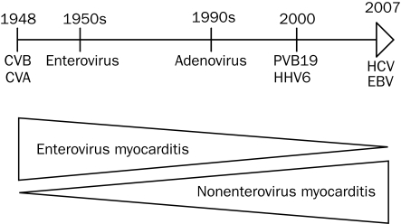
Myocarditis, an inflammatory disease of heart muscle, is an important cause of dilated cardiomyopathy worldwide. Viral infection is also an important cause of myocarditis, and the spectrum of viruses known to cause myocarditis has changed in the past 2 decades. Several new diagnostic methods, such as cardiac magnetic resonance imaging, are useful for diagnosing myocarditis. Endomyocardial biopsy may be used for patients with acute dilated cardiomyopathy associated with hemodynamic compromise, those with life-threatening arrhythmia, and those whose condition does not respond to conventional supportive therapy. Important prognostic variables include the degree of left and right ventricular dysfunction, heart block, and specific histopathological forms of myocarditis. We review diagnostic and therapeutic strategies for the treatment of viral myocarditis. English-language publications in PubMed and references from relevant articles published between January 1, 1985, and August 5, 2008, were analyzed. Main keywords searched were myocarditis, dilated cardiomyopathy, endomyocardial biopsy, cardiac magnetic resonance imaging, and immunotherapy.


Figures

FIGURE 1.
FIGURE 1.
Electrocardiograms. Left, At hospital admission, sinus tachycardia at 130 beats/min, with diffuse ST-segment elevation and 1.0 mm of PR-segment depression in leads I and II. Right, Two days after admission, normal sinus rhythm at 85 beats/min and resolution of ST-segment abnormalities.
FIGURE 2.
FIGURE 2.
Echocardiograms. At hospital admission, parasternal long-axis view showing ventricular diastole (upper left) and systole (upper right) with an estimated ejection fraction of 20% and a small pericardial effusion (arrow). Two months after admission, parasternal long-axis view showing ventricular diastole (bottom left) and systole (bottom right) with an estimated ejection fraction of 55%. Ao = aorta; LA = left atrium; LV = left ventricle; RV = right ventricle.
FIGURE 3.
FIGURE 3.
Coronary angiograms of left and right coronary arteries. A and B, Normal left main coronary artery, left anterior descending artery, and left circumflex artery and their respective branches. C, Normal right coronary artery and its respective branches. D, Hemodynamic tracings of aortic (Ao) pressure and right ventricular (RV) pressure, showing right-sided systolic pressures to be one-half of systemic pressures; Ao pressure, pulmonary artery occlusive pressure (PAOP), and right atrial (RA) pressure, showing severely elevated left-sided pressures (mean, 30 mm Hg) and moderately elevated right-sided filling pressures (mean right atrial pressure, 15 mm Hg). Pulmonary arteriolar resistance ([mean pulmonary artery pressure — pulmonary artery occlusive pressure]/cardiac output) was normal at 1.47 Wood unit, suggesting that the elevated right-sided systolic pressures were secondary to left ventricular dysfunction and not intrinsic pulmonary disease. Cardiac output calculated with the Fick formula (cardiac output = stroke volume × heart rate) was normal at 5.2 L/min due to a heart rate of 135 beats/min; however, stroke volume was severely decreased at 39 mL.
FIGURE 4.
FIGURE 4.
Endomyocardial biopsy specimens. Left, Low-power view showing diffuse lymphocytic infiltration of myocardium (arrow). Right, High-power view showing lymphocytic infiltration with myocyte destruction and surrounding myocardial edema (circle).
FIGURE 5.
FIGURE 5.
Evolution of viral causes of myocarditis over time. CVA = coxsackievirus A; CVB = coxsackievirus B; EBV = Epstein-Barr virus; HCV = hepatitis C virus; HHV6 = human herpesvirus 6; PV-B19 = parvovirus B19.

References

    1. Klingel K, Stephan S, Sauter M, et al. Pathogenesis of murine enterovirus myocarditis: virus dissemination and immune cell targets. J Virol 1996;70(12):8888-8895 - PMC - PubMed
    1. Klingel K, Sauter M, Bock CT, Szalay G, Schnorr JJ, Kandolf R. Molecular pathology of inflammatory cardiomyopathy. Med Microbiol Immunol. 2004May;193(2-3):101-117 Epub 2003 Aug 14 - PubMed
    1. Fabre A, Sheppard MN. Sudden adult death syndrome and other nonischaemic causes of sudden cardiac death. Heart 2006March;92(3):316-320 Epub 2005 May 27 - PMC - PubMed
    1. Magnani JW, Danik HJ, Dec GW, Jr, DiSalvo TG. Survival in biopsy-proven myocarditis: a long-term retrospective analysis of the histopathologic, clinical, and hemodynamic predictors. Am Heart J. 2006;151(2):463-470 - PubMed
    1. Caforio AL, Calabrese F, Angelini A, et al. A prospective study of biopsy-proven myocarditis: prognostic relevance of clinical and aetiopathogenetic features at diagnosis. Eur Heart J. 2007June;28(11):1326-1333 Epub 2007 May 9 - PubMed

MeSH terms