

Soft X-ray tomography of phenotypic switching and the cellular response to antifungal peptoids in Candida albicans
- PMID: 19880740
- PMCID: PMC2780763
- DOI: 10.1073/pnas.0906145106
Soft X-ray tomography of phenotypic switching and the cellular response to antifungal peptoids in Candida albicans
Abstract
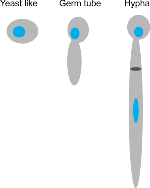
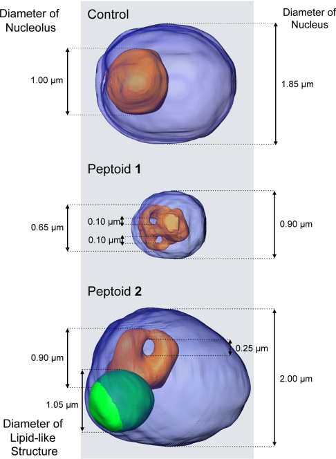
The opportunistic pathogen Candida albicans can undergo phenotypic switching between a benign, unicellular phenotype and an invasive, multicellular form that causes candidiasis. Increasingly, strains of Candida are becoming resistant to antifungal drugs, making the treatment of candidiasis difficult, especially in immunocompromised or critically ill patients. Consequently, there is a pressing need to develop new drugs that circumvent fungal drug-resistance mechanisms. In this work we used soft X-ray tomography to image the subcellular changes that occur as a consequence of both phenotypic switching and of treating C. albicans with antifungal peptoids, a class of candidate therapeutics unaffected by drug resistance mechanisms. Peptoid treatment suppressed formation of the pathogenic hyphal phenotype and resulted in striking changes in cell and organelle morphology, most dramatically in the nucleus and nucleolus, and in the number, size, and location of lipidic bodies. In particular, peptoid treatment was seen to cause the inclusion of lipidic bodies into the nucleus.
Conflict of interest statement
The authors declare no conflict of interest.
Figures


References
-
- Sudbery P, Gow N, Berman J. The distinct morphogenic states of Candida albicans. Trends Microbiol. 2004;12:317–324. - PubMed
-
- Ashman RB, et al. Innate versus adaptive immunity in Candida albicans infection. Immunol Cell Biol. 2004;82:196–204. - PubMed
-
- Monk BC, Goffeau A. Outwitting multidrug resistance to antifungals. Science. 2008;321:367–369. - PubMed
-
- Rappleyel CA, Goldman WE. Fungal stealth technology. Trends Immunol. 2008;29:18–24. - PubMed
-
- Enoch DA, Ludlam HA, Brown NM. Invasive fungal infections: A review of epidemiology and management options. J Med Microbiol. 2006;55:809–818. - PubMed
Publication types
MeSH terms
Substances
Grants and funding
LinkOut - more resources
Full Text Sources
