Review
. 2010 Feb;77(2):111-25.
doi: 10.1124/mol.109.061234. Epub 2009 Oct 30.

Structure and function of heterotrimeric G protein-regulated Rho guanine nucleotide exchange factors

Affiliations
Review

Structure and function of heterotrimeric G protein-regulated Rho guanine nucleotide exchange factors

Mohamed Aittaleb et al. Mol Pharmacol. 2010 Feb.

Abstract

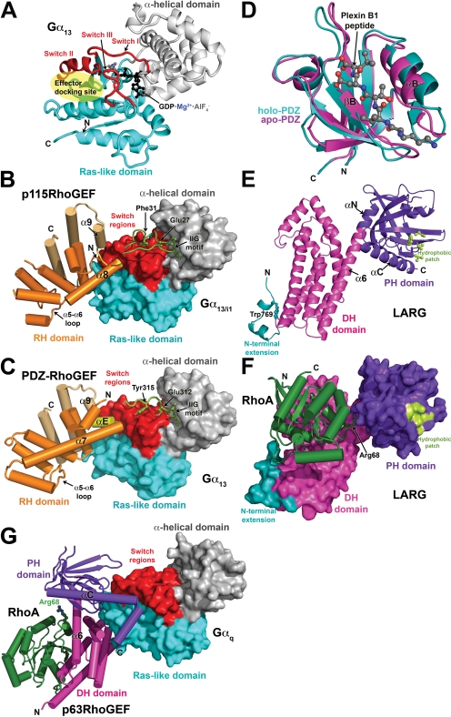
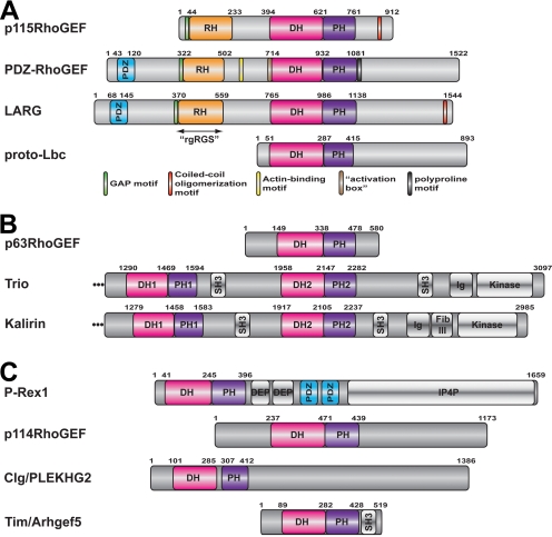
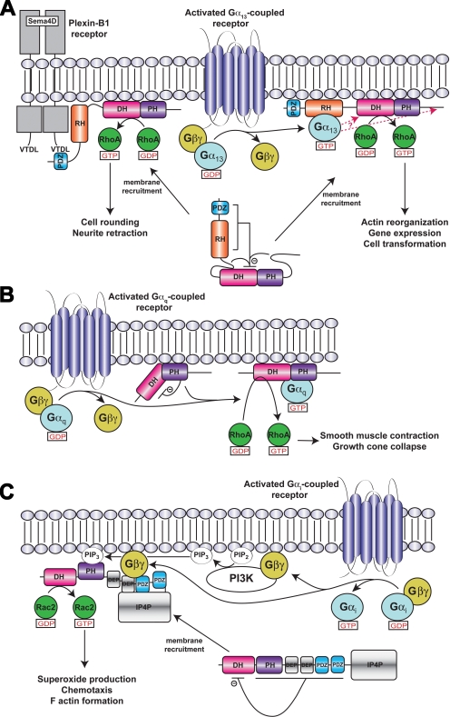
Activation of certain classes of G protein-coupled receptors (GPCRs) can lead to alterations in the actin cytoskeleton, gene transcription, cell transformation, and other processes that are known to be regulated by Rho family small-molecular-weight GTPases. Although these responses can occur indirectly via cross-talk from canonical heterotrimeric G protein cascades, it has recently been demonstrated that Dbl family Rho guanine nucleotide exchange factors (RhoGEFs) can serve as the direct downstream effectors of heterotrimeric G proteins. Heterotrimeric Galpha(12/13), Galpha(q), and Gbetagamma subunits are each now known to directly bind and regulate RhoGEFs. Atomic structures have recently been determined for several of these RhoGEFs and their G protein complexes, providing fresh insight into the molecular mechanisms of signal transduction between GPCRs and small molecular weight G proteins. This review covers what is currently known about the structure, function, and regulation of these recently recognized effectors of heterotrimeric G proteins.


Figures

Fig. 1.
Fig. 1.
Structural studies of heterotrimeric G protein-regulated RhoGEFs. a, structure of Gα13 in its active, GDP-AlF4-bound conformation. The Ras-like domain is colored cyan, and the α-helical domain is gray. The three nucleotide-dependent switch regions (switch I–III) are red. The canonical effector docking site, a shallow canyon formed between switch II and the α3 helix, is indicated by the transparent yellow ellipse. b, the p115RhoGEF rgRGS domain in complex with Gα13/i1·GDP·AlF4. The Gα subunit is shown as a molecular surface and is colored as in a. The p115RhoGEF RH domain (orange) binds to the effector docking site of the Gα13/i1 chimera with its α8-α9 loop (yellow). Noncanonical helices that extend the C terminus of the RH domain are colored in gold. The N-terminal motif responsible for GAP activity (green) binds to the α-helical domain with its IIG motif, followed by residues that stabilize catalytic residues in the GTPase active site. c, the rgRGS domain of PDZ-RhoGEF in complex with Gα13·GDP·AlF4. A short α helix (αE, colored yellow) binds in the effector docking site, which is expanded relative to the effector docking site in the Gα13/i1-p115RhoGEF complex. The N-terminal motif follows the same path as in the complex with p115RhoGEF, but there is no productive interaction with catalytic residues, explaining the absence of GAP activity. d, the LARG PDZ domain. The plexin B1 C-terminal octapeptide binds in a cleft formed between the βB strand and the αB helix, only the last five residues of the peptide being well defined. Ligand binding induces an atypical conformational change in the domain that expands the ligand binding cleft, particularly at the base of the domain (toward bottom of panel). e, the structure of the tandem DH/PH domains of LARG in their RhoA-bound conformation. Elements characteristic of the Lbc subfamily (Fig. 2a) include an N-terminal extension (teal) that coalesces around a buried tryptophan side chain (Trp769) and a hydrophobic patch on the surface of the PH domain (light green) that was recently shown to be critical for RhoA signaling in cells (Aittaleb et al., 2009). The α6 helix of the DH domain is fused to the αN helix of the PH domain to create a continuous, flexible helix that links the two domains. Residues in the α6-αN linker region form critical contacts with switch II of RhoA. f, the LARG DH/PH-RhoA complex. The tandem DH/PH domain is shown as a molecular surface and is colored as in e. RhoA binds principally to the DH domain, although the PH domain also contributes a small amount of buried surface area. Arg68 from switch II of RhoA (ball and stick model) interacts with residues in the α6-αN hinge. The complex is oriented such that the expected plane of the membrane is along the top of the panel. g, atomic structure of the Gαq-p63RhoGEF-RhoA complex. Gαq is shown as a molecular surface, and its domains are colored as for Gα13 in a. The DH (magenta) and PH (purple) domains both contact Gαq, the extended αC helix of the PH domain binding in the effector docking site. Nucleotide-free RhoA binds on the opposite surface of the DH domain from Gαq.
Fig. 2.
Fig. 2.
Domain structure of RhoGEFs reported to be directly regulated by heterotrimeric G proteins. The amino acid numbers shown above each protein correspond to the human ortholog. For domains that have not been structurally characterized, domain boundaries were assigned based on sequence alignment with their closest homologs of known structure unless otherwise indicated. a, RhoGEFs regulated by Gα12/13 subunits. p115RhoGEF, LARG, and PDZ-RhoGEF constitute the RH-RhoGEF subfamily and are characterized by an N-terminal RH domain. Short sequence motifs that seem to play functional roles are shown as small bars and are color-coded according to the key at the bottom of the panel. b, RhoGEFs regulated by Gαq subunits. All three RhoGEFs have closely related DH/PH domains that constitute the TrioC subfamily. c, RhoGEFs regulated by Gβγ subunits. None of these RhoGEFs are closely related, although p114RhoGEF belongs to the Lbc subfamily of RhoGEFs; hence, its DH/PH domains are closely related to those of the RH-RhoGEFs and Lbc. The boundaries for the PDZ domains and the domains shown in gray were assigned based on the GenPept entry for each protein.
Fig. 3.
Fig. 3.
Models of RhoGEF activation. a, LARG/PDZ-RhoGEF activation. Membrane recruitment and RhoA activation is mediated by receptors or other proteins that interact with the PDZ domain, such as the plexin B1 receptor, or by activated Gα13 subunits, which interact with the RH domain. Plexin B1 signaling to RhoA does not seem to require Gα13 (Perrot et al., 2002). Although membrane recruitment is one aspect of signal transduction to RH-RhoGEFs, additional interactions at the cell membrane involving the various structural domains and sequence motifs of the RH-RhoGEF are probably required for full activation (Bhattacharyya and Wedegaertner, 2003; Bhattacharyya et al., 2009). Indeed, there is evidence that Gα13 interacts with other regions in addition to the RH domain (indicated by red arrows) to stabilize a high-affinity complex and stimulate GEF activity (Suzuki et al., 2009b). Autoinhibition of basal activity is believed to be imposed by the cooperative actions of the N-terminal region and C-terminal tail of the RhoGEF, as well as by an acidic sequence in the RH-DH linker region (Zheng et al., 2009). Not included in this schematic is the hydrophobic patch of the PH domain, which is required for RhoA activation in cells and probably mediates protein-protein interactions at the membrane surface (Aittaleb et al., 2009). b, p63RhoGEF/TrioC subfamily activation. Unlike in RH-RhoGEFs, p63RhoGEF seems to be membrane-associated and the PH domain of p63RhoGEF autoinhibits basal activity, in part because of unfavorable interactions between switch II of RhoA and residues in the α6-αN linker region of p63RhoGEF. The bridging interactions of the Gα subunit are proposed not only to fix the PH domain in a less inhibitory conformation but also to activate the DH domain allosterically. c, Activation of P-Rex1 by Gβγ subunits in neutrophils. In neutrophils, P-Rex1 is a Rac2-specific GEF (Dong et al., 2005; Welch et al., 2005) that can be fully activated by GPCRs because of the presence of class IB PI3K (Donald et al., 2004), which are likewise activated by Gβγ subunits. The mechanism of activation shown is based on that proposed by Urano et al. (2008) with Gβγ binding to an intramolecular complex formed by the second DEP, first PDZ, and IP4P domains. Binding to Gβγ is expected to relieve the inhibition imposed by the DEP and PDZ domains (Hill et al., 2005) and recruit the enzyme to the cell surface.

References

    1. Aghazadeh B, Lowry WE, Huang XY, Rosen MK. (2000) Structural basis for relief of autoinhibition of the Dbl homology domain of proto-oncogene Vav by tyrosine phosphorylation. Cell 102: 625–633 - PubMed
    1. Aghazadeh B, Zhu K, Kubiseski TJ, Liu GA, Pawson T, Zheng Y, Rosen MK. (1998) Structure and mutagenesis of the Dbl homology domain. Nat Struct Biol 5: 1098–1107 - PubMed
    1. Aittaleb M, Gao G, Evelyn CR, Neubig RR, Tesmer JJ. (2009) A conserved hydrophobic surface of the LARG pleckstrin homology domain is critical for RhoA activation in cells. Cell Signal 21: 1569–1578 - PMC - PubMed
    1. Appert-Collin A, Cotecchia S, Nenniger-Tosato M, Pedrazzini T, Diviani D. (2007) The A-kinase anchoring protein (AKAP)-Lbc-signaling complex mediates alpha1 adrenergic receptor-induced cardiomyocyte hypertrophy. Proc Natl Acad Sci U S A 104: 10140–10145 - PMC - PubMed
    1. Aurandt J, Vikis HG, Gutkind JS, Ahn N, Guan KL. (2002) The semaphorin receptor plexin-B1 signals through a direct interaction with the Rho-specific nucleotide exchange factor, LARG. Proc Natl Acad Sci U S A 99: 12085–12090 - PMC - PubMed

Publication types

MeSH terms

Substances

LinkOut - more resources