. 2009 Oct;21(10):3379-96.
doi: 10.1105/tpc.108.064188. Epub 2009 Oct 30.

Sfp-type 4'-phosphopantetheinyl transferase is indispensable for fungal pathogenicity

Affiliations

Sfp-type 4'-phosphopantetheinyl transferase is indispensable for fungal pathogenicity

Ralf Horbach et al. Plant Cell. 2009 Oct.

Abstract

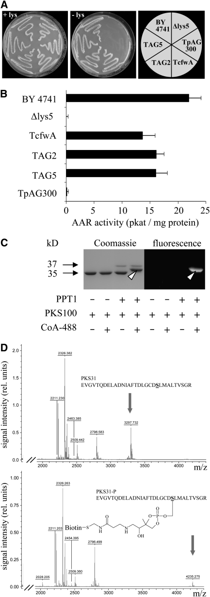
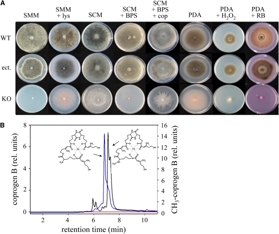
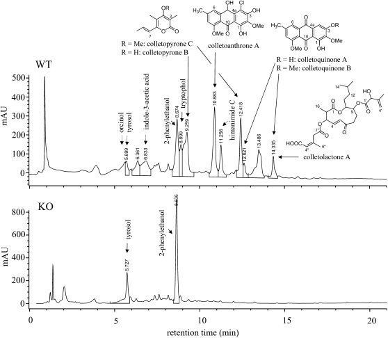
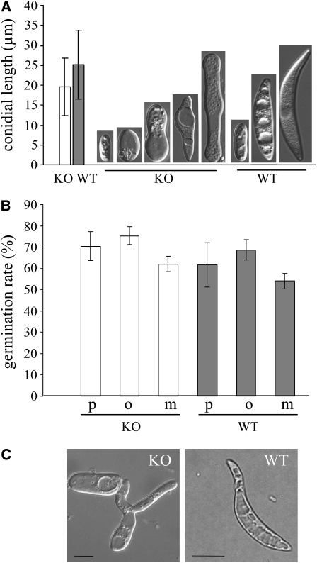
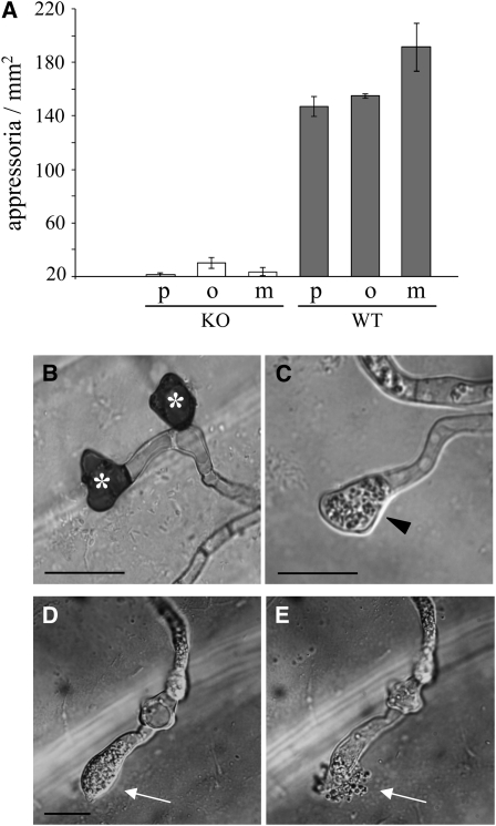
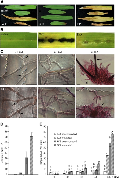
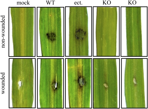
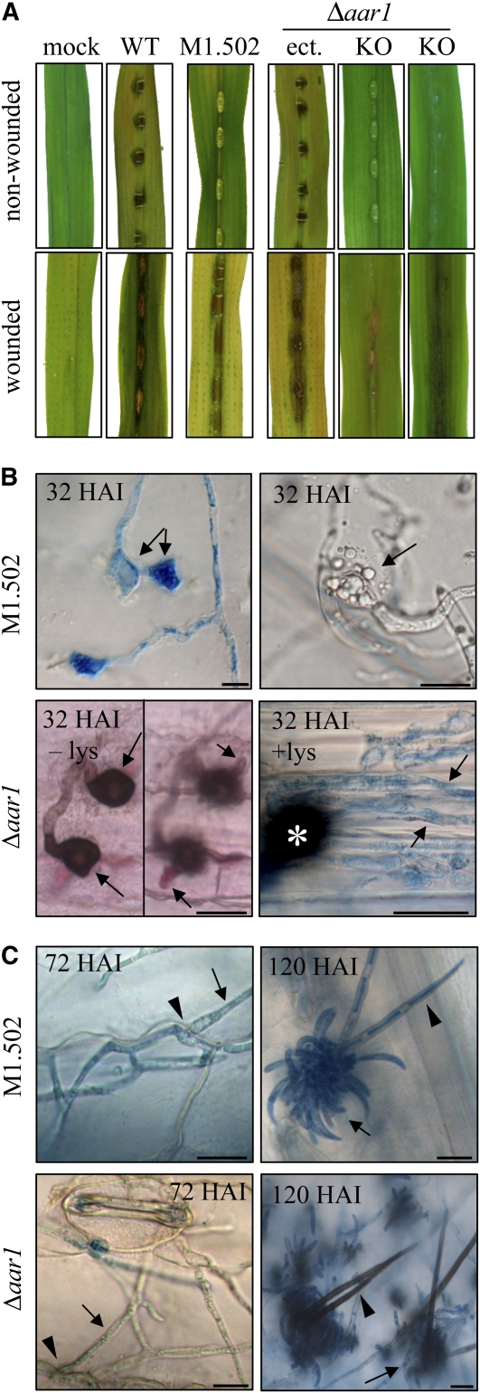
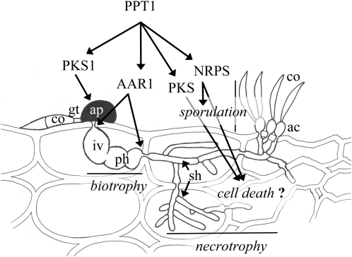
In filamentous fungi, Sfp-type 4'-phosphopantetheinyl transferases (PPTases) activate enzymes involved in primary (alpha-aminoadipate reductase [AAR]) and secondary (polyketide synthases and nonribosomal peptide synthetases) metabolism. We cloned the PPTase gene PPT1 of the maize anthracnose fungus Colletotrichum graminicola and generated PPTase-deficient mutants (Deltappt1). Deltappt1 strains were auxotrophic for Lys, unable to synthesize siderophores, hypersensitive to reactive oxygen species, and unable to synthesize polyketides (PKs). A differential analysis of secondary metabolites produced by wild-type and Deltappt1 strains led to the identification of six novel PKs. Infection-related morphogenesis was affected in Deltappt1 strains. Rarely formed appressoria of Deltappt1 strains were nonmelanized and ruptured on intact plant. The hyphae of Deltappt1 strains colonized wounded maize (Zea mays) leaves but failed to generate necrotic anthracnose disease symptoms and were defective in asexual sporulation. To analyze the pleiotropic pathogenicity phenotype, we generated AAR-deficient mutants (Deltaaar1) and employed a melanin-deficient mutant (M1.502). Results indicated that PPT1 activates enzymes required at defined stages of infection. Melanization is required for cell wall rigidity and appressorium function, and Lys supplied by the AAR1 pathway is essential for necrotrophic development. As PPTase-deficient mutants of Magnaporthe oryzea were also nonpathogenic, we conclude that PPTases represent a novel fungal pathogenicity factor.


Figures

Figure 1.
Figure 1.
Cloning of the C. graminicola PPTase Gene by Complementation of the S. cerevisiae Δlys5 Mutant, Confirmation of AAR Activity, and 4′-Phosphopantetheinylation of a PKS Fragment. (A) The reference yeast strain BY4741 and Δlys5 transformants TcfwA, TAG2, and TAG5 expressing PPTase cDNAs from A. nidulans and C. graminicola, respectively, were able to grow on medium lacking Lys (-lys). By contrast, Δlys5 or the Δlys5 transformant containing the empty expression vector (TpAG300) were auxotroph for Lys. All yeast strains grew on medium amended with Lys (+lys). (B) AAR activity was detectable in cell-free extracts from strains BY4741, TcfwA, TAG2, and TAG5 but not in extracts from the Δlys5 strain and TpAG300. Three independent experiments were performed; bars represent sd. (C) 4′-phosphopantetheinylation of a 100–amino acid C. graminicola PKS1 fragment (PKS100) fused to GST, as indicated by a band shift in Coomassie blue–stained gels and by fluorescence (arrowheads), only occurred when affinity-purified PPT1 and PKS100 and fluorochrome ATTO488-conjugated CoA were present in the reaction mixture. (D) Affinity-purified PKS100 was incubated with PPT1 in the absence (top) or presence (bottom) of biotin-conjugated CoA. After tryptic digest, MALDI-TOF MS analysis of a 31–amino acid peptide (PKS31) containing the conserved DSL phosphopantetheinylation site showed a mass shift only when biotin-conjugated CoA was present in the reaction mixture. Arrows indicate peaks corresponding to PKS31 (calculated m/z = 3299.645, measured m/z = 3297.732, error of 0.058%) and the phosphopantetheinylated PKS31-P (calculated m/z = 4237.705, measured m/z = 4235.275, error of 0.057%).
Figure 2.
Figure 2.
Growth Assays and Formation of Siderophores. (A) In comparison with the wild-type strain and a strain carrying the ectopically integrated knockout vector (ect.), the KO strain is unable to synthesize melanin or to grow under iron-depleting conditions and is hypersensitive to reactive oxygen species. SMM, synthetic minimal medium; SCM, synthetic complete medium; BPS, bathophenanthroline-disulfonate; cop, desferri-coprogen; PDA, potato dextrose agar; RB, rose bengal. (B) Analysis of siderophores formed by wild-type and Δppt1 strains. HPLC profiles of culture filtrates indicate that the wild-type strain secretes coprogen B (blue line) and 2-N-methylcoprogen B (black line) into the culture medium. Δppt1 strains do not secrete any siderophores (red line).
Figure 3.
Figure 3.
HPLC Profiles and Secondary Metabolites Identified in Culture Filtrates of Wild-Type and Δppt1 (KO) Strains. Chemical structures of orcinol, tryptophol, colletopyrone B and C, colletoanthrone A, colletoquinone A and B, and colletolactone A were identified by NMR. Tyrosol, indole-3-acetate, phenylethanol, and himanimide C were identified by employing reference data (HPLC retention time, UV, and mass spectra) from the IBWF compound library. The detection wavelength was 210 nm.
Figure 4.
Figure 4.
Formation of Asexual Spores. (A) Size and shape of conidia formed by wild-type and Δppt1 (KO) strains in liquid medium. Bars represent ± sd (n = 300). Conidial lengths of wild-type and KO strains differed significantly from each other (P < 0.05). (B) Comparison of germination rates of conidia of KO strains (white columns) and the wild type (gray columns) on different hydrophobic substrata. p, polyester sheets; o, onion epidermis; m, maize surface. Bars represent ± sd (n = 600, P > 0.05). (C) Germlings of a KO strain and the wild type. Note the branching of the germ tube of the KO strain. Bars = 10 μm.
Figure 5.
Figure 5.
Δppt1 (KO) Strains Are Unable to Differentiate Functional Appressoria. (A) In contrast with the wild-type strain (gray columns), KO strains (white columns) formed significantly lower numbers of appressoria on hydrophobic substrata (P < 0.05). p, polyester sheets; o, onion epidermis; m, maize surface. Three independent experiments were performed (total number of fungal structures counted = 1842). Bars represent ± sd. (B) Melanized wild-type appressoria (asterisks) formed on onion surface. (C) Appressoria of KO strain (arrowhead) lacked melanization. (D) and (E) Bursting appressorium and cytoplasm release from a KO strain (arrows, compare [D] and [E]). Bars = 10 μm.
Figure 6.
Figure 6.
Expression of PPT1 during Pathogenic Development of C. graminicola. Expression of the PPT1:eGFP fusion in infected maize (top row) and onion (bottom row). By 24 HAI, conidia (arrowheads) had formed appressoria (arrows) on the plant surface. At 32, 48, 72, and 120 HAI, infection vesicles, primary hyphae, secondary hyphae, and newly formed conidia had formed, respectively, as indicated by arrows. GFP fluorescence is visible in all infection structures formed. Bars = 10 μm.
Figure 7.
Figure 7.
Δppt1 Strains Are Nonpathogenic and Have Defects in Asexual Sporulation but Can Colonize Maize Leaf Tissue. (A) Plant infection assays showed that the wild type, but not a representative Δppt1 (KO) strain, caused anthracnose disease symptoms. Mock inoculation was performed with sterile 0.01% (v/v) Tween 20. In KO strains complemented with the PPT1 gene (CP), full virulence on intact leaves was restored. (B) Macroscopy evaluation of symptom development at 6 DAI on wounded leaves inoculated with the wild type and a KO strain; mock-inoculated leaves served as control. (C) Micrographs of wounded leaves inoculated with the wild type and a representative KO strain showing fungal in planta development over 6 DAI. Black arrows indicate hyphae penetrating anticlinal plant cell walls at 2 DAI, and the arrowhead indicates a hyphal appressorium-like structure at the site of penetration; at 4 DAI, epidermal cells were colonized by wild type and the Δppt1 strain; at 6 DAI, acervuli with conidia (arrowhead) and setae (arrow) were formed by the wild-type strain, whereas development of the KO strain was terminated when initials of acervuli (arrow) had formed. Samples were stained with acid fuchsin. Bars = 10 μm. (D) Relative DNA amounts of the GPD1 gene from wild-type and KO strains in wounded and nonwounded leaves as determined by quantitative PCR at five time points. Letters indicate significance groups, and bars indicate ± sd (n = 3). (E) Number of conidia produced by wild-type and KO strains on wounded and nonwounded leaves at 6 DAI. Patterns of columns are as in (D). Bars indicate ± sd (n = 80, P < 0.05).
Figure 8.
Figure 8.
Δppt1 Strains of M. oryzae Are Nonpathogenic on Rice. Plant infection assays showed that the wild-type strain and a strain with an ectopic integration of the KO construct (ect.) infected and caused symptoms on wounded and nonwounded leaves 7 DAI. The ΔMoppt1 (KO) strains were unable to cause disease symptoms on wounded or on nonwounded leaves. Representative photographs are shown. Three independent experiments were performed.
Figure 9.
Figure 9.
Synthesis of Melanin and Lys Is Required for Pathogenicity. (A) Plant infection assays showed that the wild type, but not the melanin-deficient strain M1.502 or the Lys-deficient Δaar1 strain (KO), caused anthracnose disease symptoms on nonwounded maize leaves. Symptom development was restored on wounded leaves; a strain with an ectopic integration of the AAR1 KO construct and mock-inoculated leaves served as controls. (B) Micrographs of intact leaves inoculated with M1.502 showed functionally aberrant appressoria on the intact plant surface, as indicated by lateral germination (32 HAI, left image, arrows) or ruptured appressoria (32 HAI, right image, arrow). Δaar1 strains formed melanized appressoria (32 HAI, – lys, left image, arrows) and primary infection hyphae (32 HAI, – lys, right image, arrows), which were unable to form secondary hyphae and to switch to necrotrophic development. Addition of Lys restored the ability to switch to necrotrophy, as indicated by the occurrence of thin intracellular secondary hyphae (32 HAI, + lys, arrows; asterisk marks appressorium). All samples, except Δaar1, 32 HAI, – lys, which was acid fuchsin-stained, were aniline blue stained. Bars = 10 μm. (C) On wounded leaves, M1.502 and Δaar1 strains formed secondary hyphae (72 HAI, arrows) penetrating anticlinal cell walls (72 HAI, arrowhead). Asexual sporulation occurred in both strains (120 HAI, arrows). In contrast with melanized setae of strain Δaar1 (120 HAI, arrowhead), setae of strain M1.502 were nonmelanized (120 HAI, arrowhead). Samples were stained with aniline blue. Bars = 10 μm.
Figure 10.
Figure 10.
4′-Phosphopantetheinyl Transferase (PPT1) Is a Central Regulator of Pathogenicity of C. graminicola. All PKSs, NRPSs, and AAR1 require activation by PPT1. Appressoria of melanin-deficient mutants are unable to invade the host, and AAR-deficient mutants show reduced penetration competence and form biotrophic infection structures but are unable to switch to a necrotrophic lifestyle. PPT1-deficient mutants are unable to kill the host cells, possibly due to their inability to synthesize toxic PKS- and/or NRPS-dependent secondary metabolites. PPT1-deficient mutants are also unable to sporulate, probably due to their inability to synthesize siderophores by NRPSs. ac, acervulus; ap, appressorium; co, conidium; gt, germ tube; iv, infection vesicle; ph, primary hypha; sh, secondary hypha. Artwork not to scale.

Similar articles

Cited by

References

    1. Antelo, L., Hof, C., Welzel, K., Eisfeld, K., Sterner, O., and Anke, H. (2006). Siderophores produced by Magnaporthe grisea in the presence and absence of iron. Z. Naturforsch. C 61 461–464. - PubMed
    1. Baker, S.E., Kroken, S., Inderbitzin, P., Asvarak, T., Li, B.Y., Shi, L., Yoder, O.C., and Turgeon, B.G. (2006). Two polyketide synthase-encoding genes are required for biosynthesis of the polyketide virulence factor, T-toxin, by Cochliobolus heterostrophus. Mol. Plant Microbe Interact. 19 139–149. - PubMed
    1. Bechinger, C., Giebel, K.-F., Schnell, M., Leiderer, P., Deising, H.B., and Bastmeyer, M. (1999). Optical measurements of invasive forces exerted by appressoria of a plant pathogenic fungus. Science 285 1896–1899. - PubMed
    1. Becker, D.M., and Lundblad, V. (2001). Introduction of DNA into yeast cells. In Current Protocols in Molecular Biology, F.M. Ausubel, R. Brent, R.E. Kingston, D.D. Moore, J.G. Seidman, J.A. Smith, and K. Struhl, eds (New York: John Wiley & Sons), pp. 13.17.11–13.17.10. - PubMed
    1. Bergstrom, G.C., and Nicholson, R.L. (1999). The biology of corn anthracnose. Knowledge to exploit for improved management. Plant Dis. 83 596–608. - PubMed

Publication types

MeSH terms

Substances

Associated data