Comparative Study
. 2010 Jan;3(1):157-64.
doi: 10.1161/CIRCHEARTFAILURE.109.899732. Epub 2009 Oct 30.

The cytoprotective effects of tumor necrosis factor are conveyed through tumor necrosis factor receptor-associated factor 2 in the heart

Affiliations
Comparative Study

The cytoprotective effects of tumor necrosis factor are conveyed through tumor necrosis factor receptor-associated factor 2 in the heart

Jana S Burchfield et al. Circ Heart Fail. 2010 Jan.

Abstract

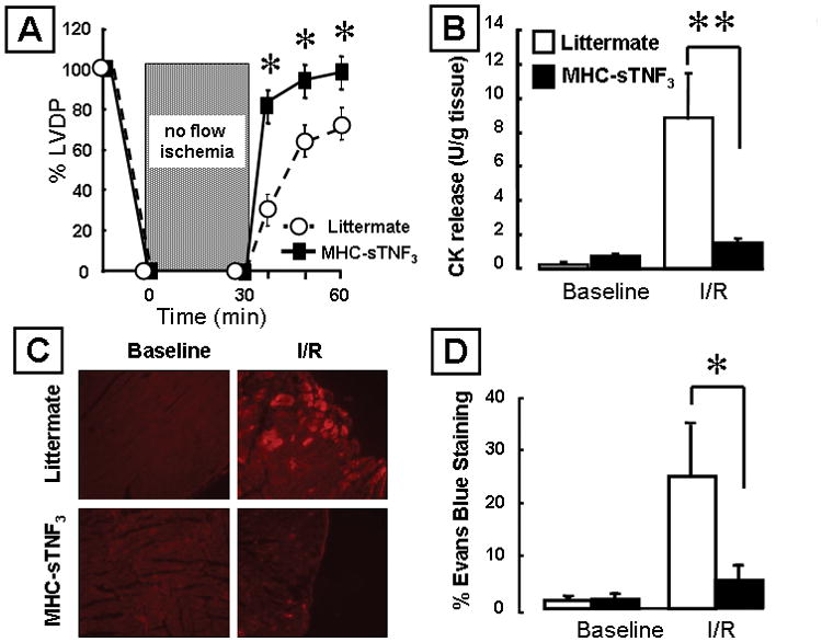
Background: Activation of both type 1 and type 2 tumor necrosis factor (TNF) receptors (TNFR1 and TNFR2) confers cytoprotection in cardiac myocytes. Noting that the scaffolding protein TNF receptor-associated factor 2 (TRAF2) is common to both TNF receptors, we hypothesized that the cytoprotective responses of TNF were mediated through TRAF2.

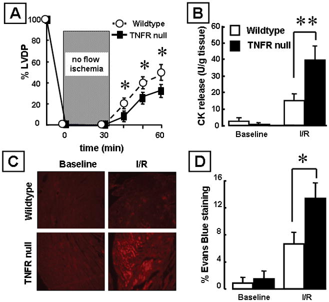
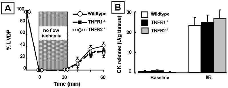
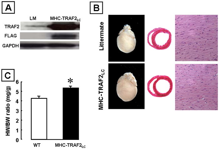
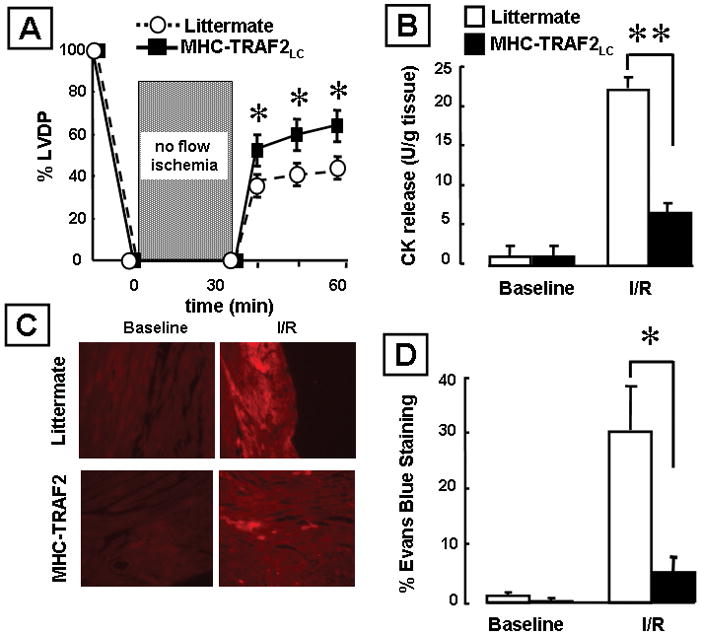
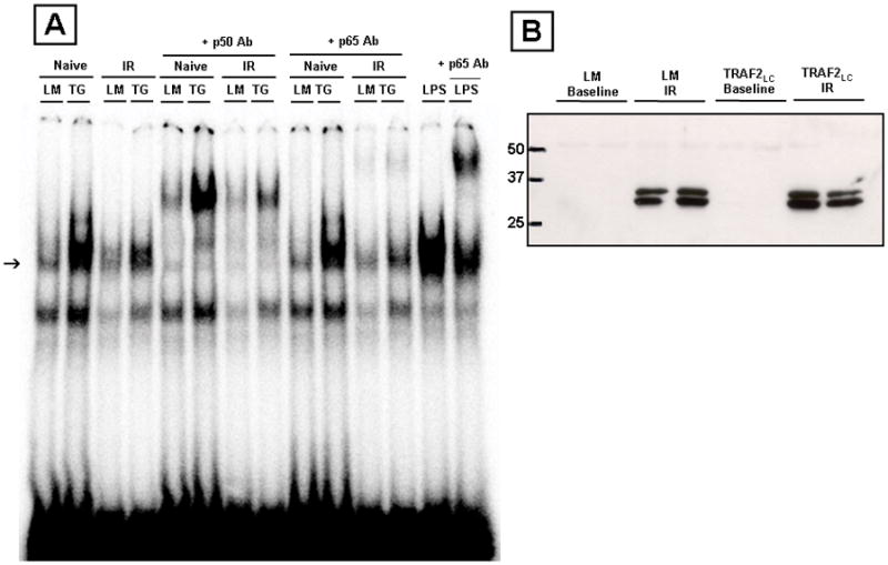
Methods and results: Mice with cardiac-restricted overexpression of low levels of TNF (MHCsTNF(3)) and TRAF2 (MHC-TRAF2(LC)) and mice lacking TNFR1, TNFR2, and TNFR1/TNFR2 were subjected to ischemia (30 minutes) reperfusion (30 minutes) injury ex vivo using a Langendorff apparatus. MHCsTNF(3) mice were protected against ischemia-reperfusion injury as shown by a significant approximately 30% greater left ventricular developed pressure, approximately 80% lower creatine kinase release, and Evans blue dye uptake compared with littermates. The extent of ischemia-reperfusion induced injury was similar in wild-type, TNFR1, and TNFR2 deficient mice; however, mice lacking TNFR1/TNFR2 had a significant approximately 40% lower left ventricular developed pressure, a approximately 65% greater creatine kinase release, and approximately 40% greater Evans blue dye uptake compared with littermates. Interestingly, MHC-TRAF2(LC) mice had a significant approximately 50% lower left ventricular developed pressure, a approximately 70% lower creatine kinase release, and approximately 80% lower Evans blue dye uptake compared with littermate controls after ischemia-reperfusion injury. Biochemical analysis of the MHC-TRAF2(LC) hearts showed that there was activation of nuclear factor-kappaB but not c-Jun N-terminal kinase activation.

Conclusions: Taken together, these results suggest that TNF confers cytoprotection in the heart through TRAF2-mediated activation of nuclear factor-kappaB.


Figures

Figure 1
Figure 1
Effects of ischemia reperfusion (I/R) injury in mice over-expressing TNF in the heart (MHC-sTNF3) and littermate control hearts. (A) Percent developed left ventricular pressure (% LVDP) after I/R injury (n=5 hearts/group). (B) Creatine kinase (CK) release in the effluent at baseline and 30 minutes after I/R injury (n= 3–5 hearts/group). (C) Representative images of Evans blue dye uptake. The red coloration indicates uptake of Evans blue dye into necrotic/permeable cardiac myocytes. (D) Group data for Evans blue dye uptake at baseline and 30 minutes after I/R injury (n= 6 hearts/group). (Key *p< 0.05 and **p < 0.005 compared to littermate controls)
Figure 2
Figure 2
Effects of ischemia reperfusion (I/R) injury in mice null for both TNF receptors (TNFR1−/−/TNFR2−/−) and wild-type control hearts. (A) Percent developed left ventricular pressure (% LVDP) after I/R injury (n= 6 hearts/group). (B) Creatine kinase (CK) release in the effluent at baseline and 30 minutes after I/R injury (n= 5 hearts/group). (C) Representative images of Evans blue dye uptake. (D) Group data for Evans blue dye uptake at baseline and 30 minutes after I/R injury (n= 6 hearts/group). (Key *p< 0.05 and **p < 0.005 compared to littermate controls)
Figure 3
Figure 3
Effects of ischemia reperfusion (I/R) injury in mice lacking TNF receptor 1 (TNFR1−/−), TNF receptor 2 (TNFR2−/−) and wild-type control hearts. (A) Percent developed left ventricular pressure (% LVDP) after I/R injury (n= 6 hearts/group). (B) Creatine kinase (CK) release in the effluent at baseline and 30 minutes after I/R injury (n= 5 hearts/group).
Figure 4
Figure 4
Characterization of transgenic mice expressing low levels of TRAF2 (MHC-TRAF2LC) in the cardiac compartment compared to littermate control mice. (A) Western blot analysis of hearts over-expressing flag tagged TRAF2. (B) Representative photographs of whole hearts and hematoxylin-and-eosin stained cross sections at the level of the papillary muscles and myocardial sections (x400). (C) Heart weight to body weight ratios (HW/BW) ratios. (Key *p < 0.005 compared to littermate controls)
Figure 5
Figure 5
Effects of ischemia reperfusion (I/R) injury in transgenic mice expressing low levels of TRAF2 (MHC-TRAF2LC) and littermate controls. Effects of ischemia reperfusion (I/R) injury in mice over-expressing TNF in the cardiac compartment (MHC-sTNF3) and littermate control hearts. (A) Percent developed left ventricular pressure (% LVDP) after I/R injury (n= 6 hearts/group). (B) Creatine kinase (CK) release in the effluent at baseline and 30 minutes after I/R injury (n= 6 hearts/group). (C) Representative images of Evans blue dye uptake. (D) Group data for Evans blue dye uptake at 30 minutes at baseline and 30 minutes after I/R injury (n= 6 hearts/group). (Key *p< 0.05 and **p < 0.005 compared to littermate controls)
Figure 6
Figure 6
NF-κB and JNK activation in transgenic mice expressing low levels of TRAF2 (MHC-TRAF2LC) and littermate control mice. (A) Representative electromobility shift assay of NF-κB binding at baseline and after I/R injury (arrow). To determine the specificity of DNA-protein binding, nuclear extracts from TRAF2LC hearts were treated with a 20X excess of unlabelled oligonucleotides, as well as by supershift assays using polyclonal antibodies directed against the p50 and p65 components of NF-κB. The positive control for the p65 supershift was obtained from a nuclear extract from a heart of a wild-type mouse that was treated with LPS at 20mg/kg for 1 hour. (B) Representative JNK activity assay in littermate and TRAF2LC hearts (12 weeks) at baseline and after I/R injury.

References

    1. Mann DL. Stress-activated cytokines and the heart: from adaptation to maladaptation. Annu Rev Physiol. 3 A.D;65:81–101. - PubMed
    1. Nakano M, Knowlton AA, Dibbs Z, Mann DL. Tumor necrosis factor-α confers resistance to injury induced by hypoxic injury in the adult mammalian cardiac myocyte. Circulation. 1998;97:1392–1400. - PubMed
    1. Eddy LJ, Goeddel DV, Wong GHW. Tumor necrosis factor-α pretreatment is protective in a rat model of myocardial ischemia-reperfusion injury. Biochem Biophys Res Commun. 1992;184:1056–1059. - PubMed
    1. Kurrelmeyer K, Michael L, Baumgarten G, Taffet G, Peschon J, Sivasubramanian N, Entman ML, Mann DL. Endogenous myocardial tumor necrosis factor protects the adult cardiac myocyte against ischemic-induced apoptosis in a murine model of acute myocardial infarction. Proc Natl Acad Sci U S A. 2000;290:5456–5461. - PMC - PubMed
    1. Lecour S, Smith RM, Woodward B, Opie LH, Rochette L, Sack MN. Identification of a novel role for sphingolipid signaling in TNF alpha and ischemic preconditioning mediated cardioprotection. J Mol Cell Cardiol. 2002;34:509–518. - PubMed

Publication types

Substances