. 2009 Jul-Sep;19(3):222-31.
doi: 10.4103/0971-3026.54887.

Radiological appearances of uterine fibroids

Affiliations

Radiological appearances of uterine fibroids

Sue Wilde et al. Indian J Radiol Imaging. 2009 Jul-Sep.

Abstract

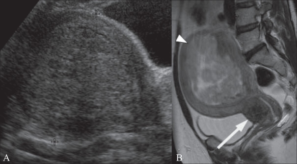
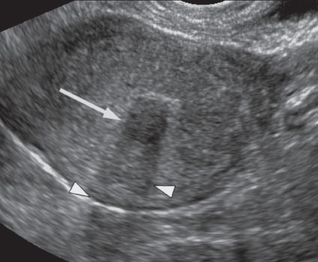
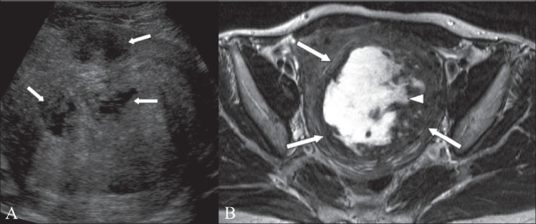
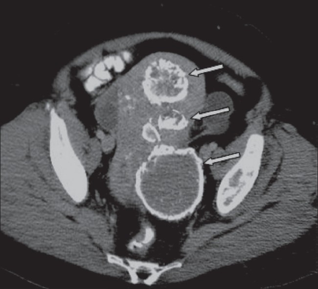
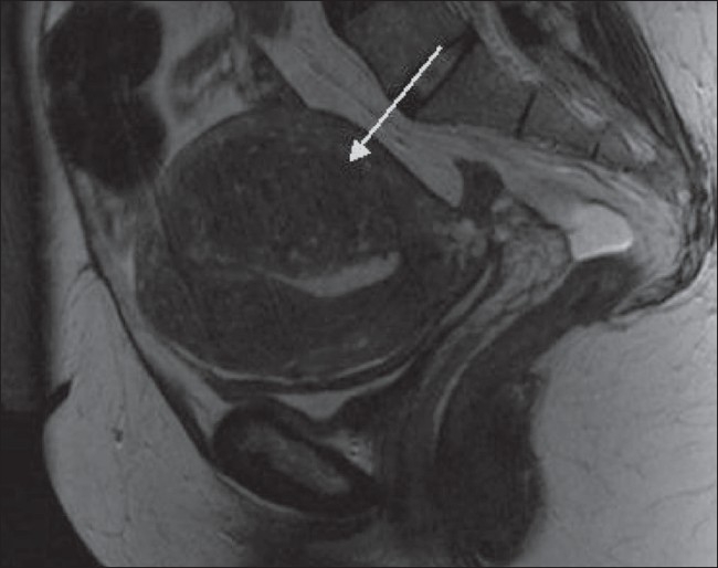
Uterine fibroids, also known as leiomyomas, are the commonest uterine neoplasms. Although benign, they can be associated with significant morbidity and are the commonest indication for hysterectomy. They are often discovered incidentally when performing imaging for other reasons. Usually first identified with USG, they can be further characterized with MRI. They are usually easily recognizable, but degenerate fibroids can have unusual appearances. In this article, we describe the appearances of typical and atypical uterine fibroids, unusual fibroid variants and fibroid mimics on different imaging modalities. Knowledge of the different appearances of fibroids on imaging is important as it enables prompt diagnosis and thereby guides treatment.


Conflict of interest statement

Conflict of Interest: None declared.

Figures

Figure 1
Figure 1
A 51-year-old woman with a history of menorrhagia. Sagittal T2W MRI shows a bulky retroverted uterus containing multiple intramural fibroids and a large submucosal fibroid (arrow) projecting into the endometrial cavity. A complex ovarian cyst is also incidentally demonstrated posterior to the uterus (arrowhead)
Figure 2
Figure 2
A 43-year-old woman with menorrhagia. Sagittal T2W MRI image shows multiple intramural fibroids (arrows); the largest (arrowhead) lying anteriorly measures 8.5 cm. These show typical low-signal intensity
Figure 3
Figure 3
A 59-year-old woman with an abdominal mass and discomfort. Sagittal T2W MRI image shows a 15-cm pedunculated, subserosal fibroid arising from the anterior uterus. There is also a small intramural fibroid lying posteriorly
Figure 4 (A, B)
Figure 4 (A, B)
A 58-year-old woman with sacral pain. A frontal pelvic radiograph (A) shows calcification overlying the left hip, initially thought to be suggestive of a chondrosarcoma. A subsequent CT scan (B) reveals an incidental calcified fibroid
Figure 5 (A, B)
Figure 5 (A, B)
A 49-year-old woman with a history of menorrhagia. Transabdominal (TA) USG image (A) shows a bulky uterus showing a 10-cm submucosal fibroid (between cursors). Sagittal T2W MRI image (B) in the same patient shows that the submucosal fibroid (arrowhead) is heterogeneous indicating degeneration. There is also a 2.5-cm cervical fibroid (arrow)
Figure 6
Figure 6
A 46-year-old woman with a history of abdominal pain. Transvaginal (TV) USG image shows a 1.1-cm submucous fibroid (arrow) with posterior acoustic shadowing (arrowheads)
Figure 7 (A, B)
Figure 7 (A, B)
A 46-year-old woman with menorrhagia. TA USG image (A) shows a 7-cm intramural fibroid containing cystic areas (arrows). Axial T2W MRI image (B) shows an 11 × 8 cm fibroid (arrows) containing central high signal, consistent with cystic degeneration (arrowhead)
Figure 8
Figure 8
A 51-year-old woman known to have fibroids, which have been treated by uterine artery embolization 18 months earlier. Axial CT scan shows several fibroids with peripheral calcification (arrows)
Figure 9
Figure 9
A 45-year-old woman with a large, asymptomatic abdominal mass. Axial CT scan image shows a 30-cm heterogeneous mass (arrowheads) that extends up to the epigastrium. The right ovarian vein is dilated (arrow) and there is mild right hydronephrosis. There is also free intra-abdominal fluid (curved arrow). Histology confirmed this to be a massive, partly degenerate fibroid
Figure 10 (A, B)
Figure 10 (A, B)
A 37-year-old woman with menorrhagia. Sagittal T2W MRI (A) shows an 18-cm, broad-based, subserosal fibroid (arrow). Repeat sagittal T2W image (B), 3 years post-UAE shows good response to treatment: the fibroid has reduced in size, now measuring 10.5 cm in AP dimension (arrow)
Figure 11
Figure 11
A 71-year-old woman shown to have uterine fibroids whilst undergoing an MRI scan of her hip. She had no gynecological symptoms. Sagittal T2W MRI image shows a 7-cm intramural fibroid (arrow), with surrounding high signal (arrowhead). Several other small intramural fibroids (curved arrows) and a trace of fluid in the pouch of Douglas are also present
Figure 12 (A, B)
Figure 12 (A, B)
A 21-year-old woman with menorrhagia and a pelvic mass. Sagittal (A) and coronal (B) T2W MRI images show a large intramural fibroid (arrows) that contains septate areas of high signal (arrowheads). The patient underwent a myomectomy and histopathology confirmed that this was a fibroid with myxoid degeneration
Figure 13 (A, B)
Figure 13 (A, B)
A 44-year-old woman, known to have uterine fibroids, admitted with acute abdominal pain and vaginal bleeding. Axial T2W (A) and T1W (B) MRI images show a heterogeneous mass (arrow) within the endometrial cavity. The mass shows peripheral high signal (arrowheads) on the T1W image, due to hemorrhage. The patient underwent a hysterectomy, and histology showed that this was a submucosal fibroid that had undergone extensive red degeneration
Figure 14
Figure 14
A 47-year-old woman with a pelvic mass. Sagittal T2W MRI image shows an enlarged heterogeneous uterus containing multiple nodules (arrows). Hysterectomy and histology showed that this was diffuse leiomyomatosis. There was no evidence of extra-uterine spread
Figure 15 (A-D)
Figure 15 (A-D)
A 65-year-old woman with a pelvic mass. Transabdominal USG image demonstrates an echogenic mass (between cursors) within the pelvis. Axial CT scan (B) shows a 10 × 15 cm mass (arrows) containing areas of fat attenuation (arrowheads). Axial T1W MRI image (C) shows that the mass (arrows) is heterogeneous, containing areas of high signal (arrowheads). Coronal fat-saturated T2W MRI image (D) confirms the presence of fat (arrows). Histological examination identified this to be a lipoleiomyoma
Figure 16 (A,B)
Figure 16 (A,B)
A 70-year-old woman with postmenopausal bleeding and a pelvic mass. Sagittal T2W MRI image (A) shows a 13.5-cm heterogeneous pelvic mass (arrows). Axial T1 MRI image (B) shows areas of high signal within the mass, including an area with a fluid-fluid level (arrow) that is in keeping with hemorrhage. The patient underwent a hysterectomy and this was confirmed to be a leiomyosarcoma
Figure 17
Figure 17
Sagittal T2W MRI image shows thickening of the junctional zone in a patient with adenomyosis. The thickening is most marked posteriorly (arrow) and several small cystic spaces can be seen within it

References

    1. Prayson RA, Hart WR. Pathologic considerations of uterine smooth muscle tumors. Obstet Gynecol Clin North Am. 1995;22:637–57. - PubMed
    1. Erdemoglu E, Kamaci M, Bayram I, Güler A, Güler SH Primary giant leiomyoma of the ovary – case report. Eur J Gynaecol Oncol. 2006;27:634–5. - PubMed
    1. Cramer SF. Patel A The frequency of uterine leiomyomas. Am J Clin Pathol. 1990;94:435–8. - PubMed
    1. Day Baird D, Dunson DB, Hill MC, Cousins D, Schectman J. High cumulative incidence of uterine leiomyoma in black and white women: Ultrasound evidence. Am J Obstet Gynecol. 2003;188:100–07. - PubMed
    1. Marshall LM, Spieglman D, Barbieri RL, Goldman MB, Manson JE, Colditz GA, Willett WC, Hunter DJ. Variation in the incidence of uterine leiomyoma among premenopausal women by age and race. Obstet Gynecol. 1997;90:967–73. - PubMed