. 2009 Nov;15(11):1298-306.
doi: 10.1038/nm.2052. Epub 2009 Nov 1.

Activation of PKC-delta and SHP-1 by hyperglycemia causes vascular cell apoptosis and diabetic retinopathy

Affiliations

Activation of PKC-delta and SHP-1 by hyperglycemia causes vascular cell apoptosis and diabetic retinopathy

Pedro Geraldes et al. Nat Med. 2009 Nov.

Abstract

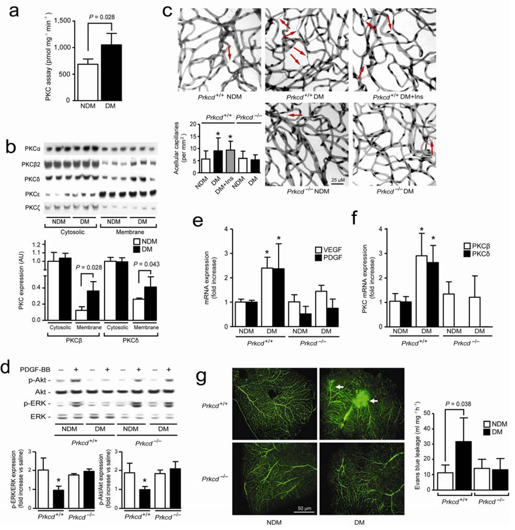
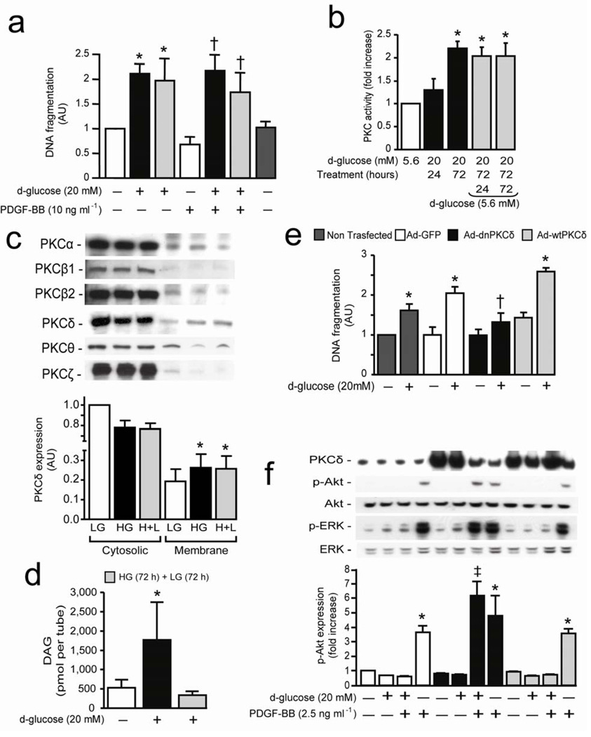
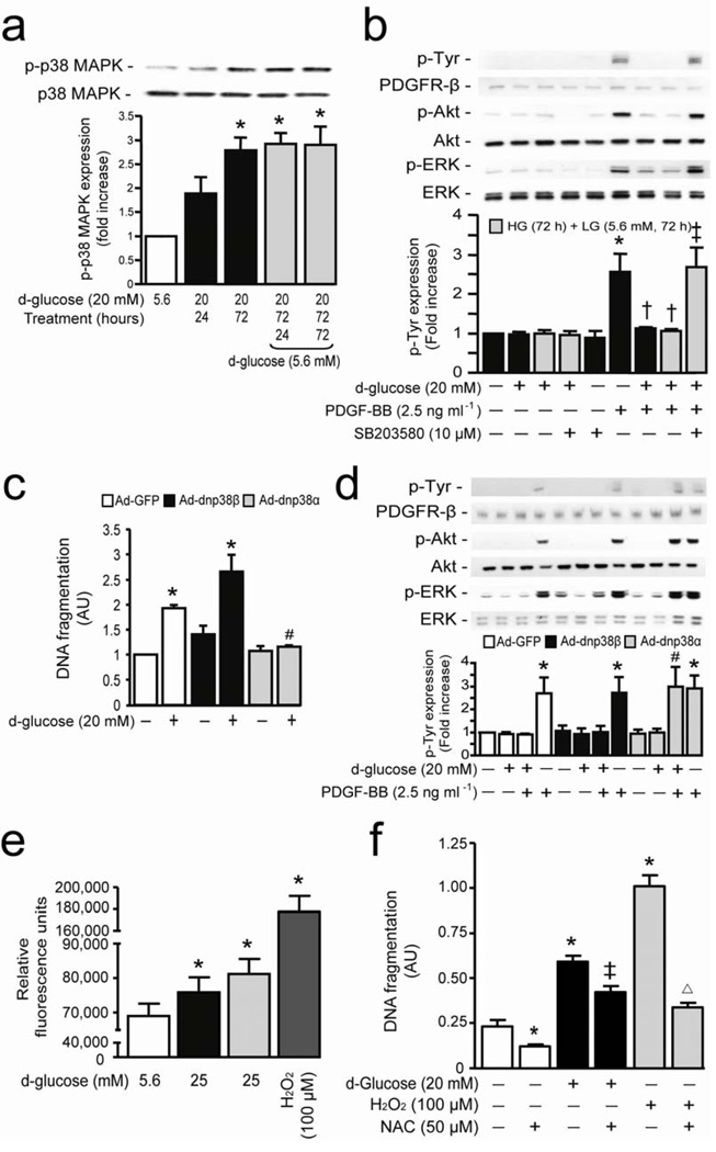
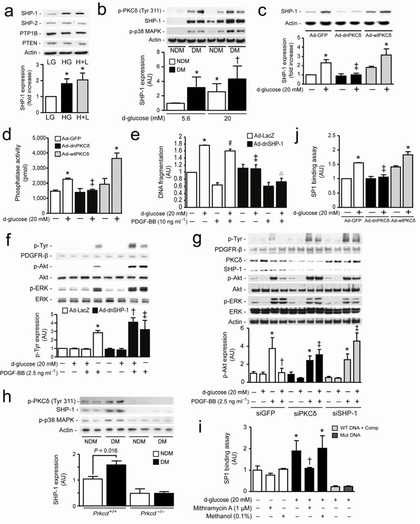
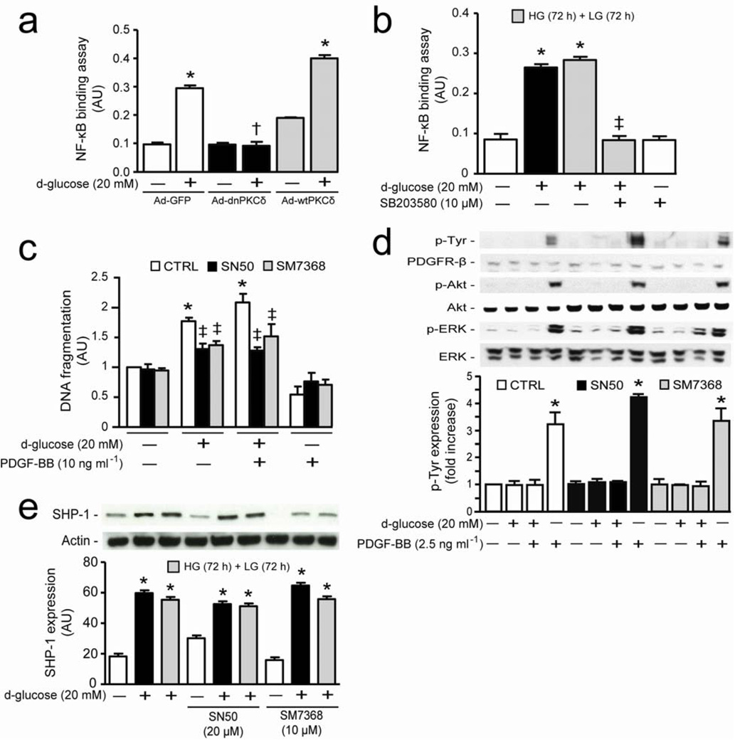
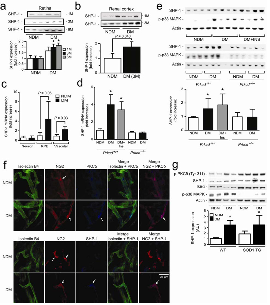
Cellular apoptosis induced by hyperglycemia occurs in many vascular cells and is crucial for the initiation of diabetic pathologies. In the retina, pericyte apoptosis and the formation of acellular capillaries, the most specific vascular pathologies attributed to hyperglycemia, is linked to the loss of platelet-derived growth factor (PDGF)-mediated survival actions owing to unknown mechanisms. Here we show that hyperglycemia persistently activates protein kinase C-delta (PKC-delta, encoded by Prkcd) and p38alpha mitogen-activated protein kinase (MAPK) to increase the expression of a previously unknown target of PKC-delta signaling, Src homology-2 domain-containing phosphatase-1 (SHP-1), a protein tyrosine phosphatase. This signaling cascade leads to PDGF receptor-beta dephosphorylation and a reduction in downstream signaling from this receptor, resulting in pericyte apoptosis independently of nuclear factor-kappaB (NF-kappaB) signaling. We observed increased PKC-delta activity and an increase in the number of acellular capillaries in diabetic mouse retinas, which were not reversible with insulin treatment that achieved normoglycemia. Unlike diabetic age-matched wild-type mice, diabetic Prkcd(-/-) mice did not show activation of p38alpha MAPK or SHP-1, inhibition of PDGF signaling in vascular cells or the presence of acellular capillaries. We also observed PKC-delta, p38alpha MAPK and SHP-1 activation in brain pericytes and in the renal cortex of diabetic mice. These findings elucidate a new signaling pathway by which hyperglycemia can induce PDGF resistance and increase vascular cell apoptosis to cause diabetic vascular complications.


Figures

Figure 1
Figure 1. PKCδ activation cause PDGF-BB and microvascular abnormalities
(a) In situ PKC activity assay was measured in retina of 3 months diabetic mice (n = 6) as compared to non-diabetic mice as described in method section. (b) Immunoblot analyses of different PKC isoform in cytosolic and membrane fraction of retinal isolated microvessels of NDM and 3 months DM mice. (c) Quantification of acellular capillaries (red arrow) of Prkcd+/+ and Prkcd−/− mouse retina (NDM Prkcd+/+, n = 22; DM Prkcd+/+, n = 19; DM+Ins Prkcd+/+, n = 12; NDM Prkcd−/−, n = 13; DM Prkcd−/−, n = 13). (d) Immunoblot analyses of intravitreous injection of saline or PDGF-BB of NDM and DM mice retina. (e) Expression of VEGF, PDGF, (f) PKCβ and PKCδ mRNA from isolated microvessels of NDM and DM Prkcd+/+ or Prkcd−/− mice. (g) Flat mount of retina infused with FITC-dextran and permeability assay using Evans blue dye. Immunoblot experiments were done at least in triplicate. Results are shown as mean ± SD of 3–4 independent experiments * P < 0.05 versus NDM Prkcd+/+ mice.
Figure 2
Figure 2. Hyperglycemia inhibits PDGF-B actions and induces pericyte apoptosis through activation of PKCδ isoform
(a) BRPC were incubated with LG (5.6 mM; white bars) or HG (20 mM; black bars) for 72 hours and then LG (gray bars) for an additional 72 hours in absence or presence of PDGF-BB. DNA fragmentation was measured according to manufacturer’s instructions. (b) In situ PKC activity assay was measured in BRPC exposed to HG or LG as described in method section. (c) Immunoblot analyses of different PKC isoform in cytosolic and membrane fraction of BPRC. (d) Total DAG was measured as described in method section. (e,f) BRPC were transfected with Ad-GFP (white bars), Ad-dnPKCδ (black bars) or Ad-wtPKCδ (gray bars). BRPC were incubated with LG or HG for 72 hours. (e) DNA fragmentation was measured according to manufacturer’s instructions. (fF) Expression of PKCδ, phospho-ERK, ERK, phospho-Akt and Akt were detected by Western blot and densitometric quantitation was measured. Results are shown as mean ± SD of 3–5 independent experiments. * P < 0.01 versus LG, † P < 0.05 versus PDGF-BB, ‡ P < 0.05 versus HG.
Figure 3
Figure 3. P38α MAPK serves as a downstream target for hyperglycemia to cause PDGF-B resistance and pericyte apoptosis
(a) BRPC were incubated with LG (5.6 mM; white bars) or HG (20 mM; black bars) for 24–72 hours and then LG (5.6 mM; gray bars) for an additional 24–72 hours. Expression of phospho-p38 MAPK and p38 MAPK were detected by Western blot. (b) Cells were exposed to LG or HG in presence or absence of SB203580 and then stimulated with PDGF-BB for 10 min. (c) BRPC were transfected with Ad-GFP (white bars), Ad-dnp38β MAPK (black bars) or Ad-dnp38α MAPK (gray bars), exposed to HG for 72 hours. DNA fragmentation was measured according to manufacturer’s instructions. (d) After transfection and exposure to LG or HG for 72 hours, cells were stimulated with PDGF-BB for 10 min. (b,d) Expression of phospho-Tyr, PDGFR-β, phospho-Akt, Akt, phospho-ERK were detected by Western blot and densitometric quantitation was measured. (e) ROS production was measured in BPRC exposed to LG, HG, H+L and H2O2 as described in the method section. (f) BPRC were exposed to LG and HG for 72 hours or H2O2 for 2 hours in absence or presence of NAC. DNA fragmentation was measured according to manufacturer’s instructions. Results are shown as mean ± SD of 3–5 independent experiments. * P < 0.05 versus LG, † P < 0.05 versus PDGF-BB, ‡ P < 0.05 versus HG, # P < 0.05 versus HG+PDGF-BB in Ad-GFP cells, Δ P < 0.05 versus H2O2.
Figure 4
Figure 4. SHP-1 causes inhibition of PDGF signaling pathway induced by hyperglycemia and PKCδ/p38α MAPK activation
(a) BRPC were exposed to LG and HG as described in the method section. Expression of SHP-1, SHP-2, PTP1B, PTEN were detected by Western blot and normalized to actin expression. (b) RtPC isolated from NDM and DM rats cultured in LG or HG for 72 hours. (c,d,j) BRPC were transfected with either ad-GFP (white bars), Ad-dnPKCδ (black bars) or Ad-wtPKCδ (grey bars) and then incubated with LG or HG for 72 hours. (d) Phosphatase activity was measured using commercially available kit as described in the method section. (e,f) Cells were transfected with either Ad-LacZ (white bars) or Ad-dnSHP-1 (black bars), exposed to HG (20 mM) for 72 hours in presence or absence of PDGF-BB. (e) Apoptosis was measured by DNA fragmentation according to manufacturer’s instructions. (g) BRPC were transfected with either GFP (white bars), PKCδ (black bars) or SHP-1 (gray bars) siRNA, exposed to LG or HG for 72 hours and then stimulated with PDGF-BB. (h) BrPC of NDM and DM Prkcd+/+ and Prkcd−/− mice were cultured in LG for 72 hours. (i,j) Transcriptional binding assay of SP1 was performed as described in the method section. (i) BRPC were exposed to LG or HG in presence or absence of mithramycin A or methanol. (b,c,f,g,h) Expression of phospho-Tyr, phospho-PKCδ, PDGFR-β, phospho-Akt, Akt, phospho-ERK, ERK, phospho-p38 MAPK, SHP-1 and actin were detected by Western blot and densitometric quantitation was measured. Results are shown as mean ± SD of 3–5 independent experiments. * P < 0.05 versus LG, † P < 0.05 versus HG or HG+LG, ‡ P < 0.05 versus HG in Ad-GFP or HG in siGFP, # P < 0.05 versus PDGF-BB, Δ P < 0.05 versus HG in Ad-dnSHP-1.
Figure 5
Figure 5. Increase of SHP-1 expression by hyperglycemia is independent of NF-κB activation
(Aa) BRPC were transfected with either ad-GFP (white bars), Ad-dnPKCδ (black bars) or Ad-wtPKCδ (gray bars) and then incubated with LG or HG for 72 hours. (b) Transcriptional binding activity assay of NF-κB was performed as described in the method section. (c) BRPC were incubated with LG or HG in absence (white bars) or presence of inhibitor or NF-κB (SN50, black bars; SM7368, gray bars). Apoptosis was measured by DNA fragmentation according to manufacturer’s instructions. (d) BRPC were incubated as described in (c) and then stimulated with PDGF-BB for 10 min. Expression of phospho-Tyr, PDGFR-β, phospho-Akt, Akt, phospho-ERK, ERK and (e) SHP-1 were detected by Western blot and densitometric quantitation was measured. Results are shown as mean ± SD of 3–5 independent experiments. * P < 0.05 versus LG, † P < 0.05 versus HG in Ad-GFP, ‡ P < 0.05 versus HG or HG+LG.
Figure 6
Figure 6. PKCδ induces SHP-1 expression and p38 MAPK activation in retina, kidney and PBMC of diabetic rodent animals
Expression of SHP-1 in (a) retina and (b) renal cortex of 1 to 6 months diabetic mice. (c) SHP-1 mRNA expression in retinal neurone, RPE and vascular of NDM and 6 month DM mice by laser capture micro-dissection as described in the method section. (d) SHP-1 mRNA, (e) SHP-1 protein expression and p38 MAPK activity in whole retina of NDM and 6 months DM Prkcd+/+ and Prkcd−/− mice. (f) Immunofluorescence of endothelial cells (isolectin B4; green), pericytes (NG2; red) and PKCδ or SHP-1 (blue) of isolated retinal microvessels from NDM and 3 months DM mice. (g) SHP-1, phospho-PKCδ, phosho-p38 MAPK, IkBα and actin expression in whole retina of NDM and DM wild-type and SOD1 transgenic mice. Immunoblot analyses and densitometric quantitation were performed. Results are shown as mean ± SD of 3–5 independent experiments. * P < 0.05 versus NDM Prkcd+/+ mice.

Comment in

References

    1. Lorenzi M, Gerhardinger C. Early cellular and molecular changes induced by diabetes in the retina. Diabetologia. 2001;44:791–804. - PubMed
    1. Isermann B, et al. Activated protein C protects against diabetic nephropathy by inhibiting endothelial and podocyte apoptosis. Nature medicine. 2007;13:1349–1358. - PubMed
    1. >The effect of intensive treatment of diabetes on the development and progression of long-term complications in insulin-dependent diabetes mellitus. The Diabetes Control and Complications Trial Research Group. N Engl J Med. 1993;329:977–986. - PubMed
    1. Mizutani M, Kern TS, Lorenzi M. Accelerated death of retinal microvascular cells in human and experimental diabetic retinopathy. J Clin Invest. 1996;97:2883–2890. - PMC - PubMed
    1. Busik JV, Mohr S, Grant MB. Hyperglycemia-induced reactive oxygen species toxicity to endothelial cells is dependent on paracrine mediators. Diabetes. 2008;57:1952–1965. - PMC - PubMed

Publication types

MeSH terms