Review
. 2009 Dec;158 Suppl 1(Suppl 1):2-13.
doi: 10.1111/j.1365-2249.2009.04022.x.

Intravenous immunoglobulins--understanding properties and mechanisms

Affiliations
Review

Intravenous immunoglobulins--understanding properties and mechanisms

A Durandy et al. Clin Exp Immunol. 2009 Dec.

Abstract

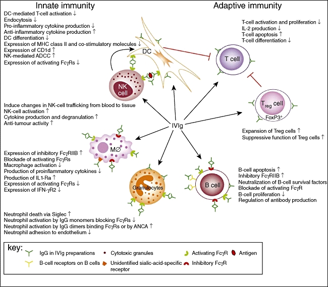
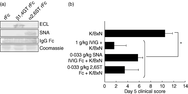
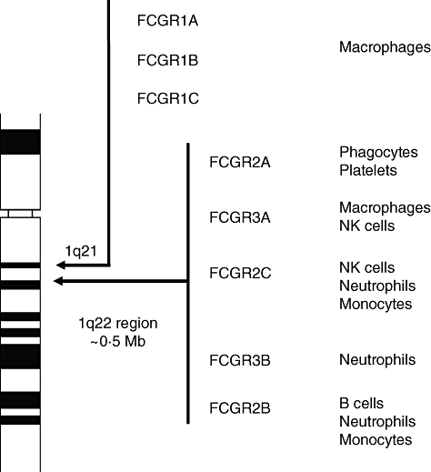
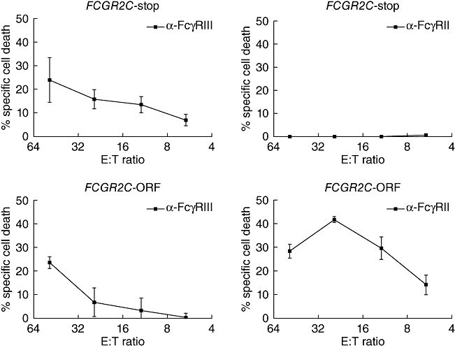
High-dose intravenous immunoglobulin (IVIg) preparations are used currently for the treatment of autoimmune or inflammatory diseases. Despite numerous studies demonstrating efficacy, the precise mode of action of IVIg remains unclear. Paradoxically, IgG can exert both pro- and anti-inflammatory activities, depending on its concentration. The proinflammatory activity of low-dose IVIg requires complement activation or binding of the Fc fragment of IgG to IgG-specific receptors (FcgammaR) on innate immune effector cells. In contrast, when administered in high concentrations, IVIg has anti-inflammatory properties. How this anti-inflammatory effect is mediated has not yet been elucidated fully, and several mutually non-exclusive mechanisms have been proposed. This paper represents the proceedings of a session entitled 'IVIg--Understanding properties and mechanisms' at the 6th International Immunoglobulin Symposium that was held in Interlaken on 26-28 March 2009. The presentations addressed how IgG may affect the cellular compartment, evidence for IVIg-mediated scavenging of complement fragments, the role of the dimeric fraction of IVIg, the anti-inflammatory properties of the minor fraction of sialylated IgG molecules, and the genetic organization and variation in FcgammaRs. These findings demonstrate the considerable progress that has been made in understanding the mechanisms of action of IVIgs, and may influence future perspectives in the field of Ig therapy.


Figures

Fig. 1
Fig. 1
A schematic representation of the proposed mechanisms of action of intravenous immunoglobulin (IVIg) on cellular immunity. IVIg targets the cellular immune compartment at multiple levels, including innate and adaptive immune cells. IVIg interacts with dendritic cells (DCs), macrophages (MØ) and granulocytes, mainly via activating and inhibitory FC gamma receptors (FcγRs). Monomeric IgG in IVIg preparations can block the interaction of immune complexes with activating FcγRs, thereby inhibiting endocytosis and phagocytosis by DCs and macrophages and activation of granulocytes. IgG dimers in IVIg preparations binding to activating FcγRs on macrophages induce the expression of the inhibitory FcγRIIB and suppress expression of interferon (IFN)-γR2, thereby inhibiting macrophage functions. In addition, IgG dimers promote antibody-dependent cell-mediated cytotoxicity (ADCC) of DCs by natural killer (NK) cells, resulting in reduced T cell activation. IgG dimers suppress macrophage and B cell functions by ligating FcγRIIB. In addition, F(ab′)2-mediated effects of natural antibodies present in IVIg have been described for DCs, granulocytes and B cells. Interactions between IVIg and regulatory T cells (Tregs) lead to expansion and increased suppressive function of Tregs. The immunological effects depicted are not mutually exclusive and are likely to work in synergy. Reprinted from Tha-In et al.[28], copyright 2008, with permission from Elsevier.
Fig. 2
Fig. 2
Attenuation of in situ C3b binding by intravenous immunoglobulin (IVIg). (a) Ischaemia/reperfusion (I/R) brain injury causes increased presence of the complement protein C3b in the affected tissue, and post-injury IVIg treatment reduces the I/R-mediated C3b binding. (b) C3b binding is strongest at the site of the lesion, i.e. in the ipsilateral region. (c) Immunoprecipitation analysis of brain samples from the ischaemic hemisphere of IVIg-treated mice, showing that human IgG binds mouse C3b. Shams: animals subjected to the same extent of anaesthesia and surgery as I/R animals, except for the middle artery occlusion; controls for the effect of surgical and anaesthesiological injury. Vehicle: animals infused with the stabilizer for the immunoglobulins in IVIg preparation; controls for the specificity of immunoglobulin effect in the IVIg preparation.
Fig. 3
Fig. 3
Factors affecting dimer formation. Larger donor pool size, low temperature, high pH and high immunoglobulin (Ig)G concentration increase the dimer content of therapeutic Ig formulations.
Fig. 4
Fig. 4
Immunofluorescent staining patterns of the different immunoglobulin (Ig) preparations analysed by incubation on human epithelial type 2 (HEP-2) cells. (a) Positive anti-centromere control serum, showing typical speckled staining pattern. Inset shows May–Grünwald–Giemsa staining of HEP-2 cells and black inset is the conjugate control. Staining of perinuclear antigens characterizes the patterns for intravenous immunoglobulin (IVIg) (b) and the monomeric IgG fraction (c). The dimeric IgG fraction shows enhanced nuclear and cytoplasmic staining (d). IgG purified from adult single-donor serum (e) and IgM from cord-blood serum (f) showed similar staining on perinuclear structures comparable to monomeric IgG and IVIg.
Fig. 5
Fig. 5
Recombinant, sialylated immunoglobulin (Ig)G Fc fragments are anti-inflammatory and comparable to native intravenous immunoglobulin (IVIg). Recombinant human IgG1 was digested with papain, and Fc fragments were purified. The recombinant Fc fragments were galactosylated and sialylated in vitro with α-2,6-sialyltransferase (α-2,6-ST). (a) Glycosylation was confirmed by lectin blotting for terminal galactose with enhanced chemiluminescence (ECL) (top) or α-2,6-sialic acid with Sambucus nigra agglutinin (SNA) (middle); Coomassie loading controls are shown (bottom). (b) Mice were administered 1 g/kg IVIg, 0·033 g/kg SNA+ IVIg Fc fragments, or 0·33 g/kg sialylated recombinant Fc (α-2,6-ST rFc) 1 h before K/BxN sera, and footpad swelling was monitored over the next several days. Means and standard deviations of clinical scores of four to five mice per group are plotted. *P < 0·05. From Anthony RM et al.[58]; reprinted with permission from the American Association for the Advancement of Science.
Fig. 6
Fig. 6
Human Fc gamma (Fcγ) receptors. In humans, the genes encoding for the Fcγ receptors are located on chromosome 1. The receptors are expressed on different cell types, as indicated on the right.
Fig. 7
Fig. 7
Redirected antibody-dependent cell-mediated cytotoxicity (rADCC) of effector natural killer (NK) cells against target cells. Peripheral blood lymphocytes were isolated and Fc gamma receptor IIC (FcγRIIC) functionality was assessed by rADCC. Cells from both FCGR2C-Stop and FCGR2C-open reading frame (ORF) genotyped donors killed anti-FcγRIII-coated targets with similar kinetics (left panels). In contrast, only cells from FCGR2C-ORF genotyped donors were capable of killing anti-FcγRII-coated targets (right panels) (n = 4). Data are expressed as mean ± standard error of the mean.

References

    1. Nimmerjahn F, Ravetch JV. Anti-inflammatory actions of intravenous immunoglobulin. Annu Rev Immunol. 2008;26:513–33. - PubMed
    1. Conley ME, Dobbs AK, Farmer DM, et al. Primary B cell immunodeficiencies: comparisons and contrasts. Annu Rev Immunol. 2009;27:199–227. - PubMed
    1. Durandy A, Wahn V, Petteway S, Gelfand EW. Immunoglobulin replacement therapy in primary antibody deficiency diseases – maximizing success. Int Arch Allergy Immunol. 2005;136:217–29. - PubMed
    1. Gürcan HM, Ahmed AR. Efficacy of various intravenous immunoglobulin therapy protocols in autoimmune and chronic inflammatory disorders. Ann Pharmacother. 2007;41:812–23. - PubMed
    1. Kazatchkine MD, Kaveri SV. Immunomodulation of autoimmune and inflammatory diseases with intravenous immune globulin. N Engl J Med. 2001;345:747–55. - PubMed

Publication types