. 2009 Mar;152(1):157-66.
doi: 10.1016/j.jss.2008.06.029. Epub 2008 Jul 26.

Endothelial differentiation of adipose-derived stem cells: effects of endothelial cell growth supplement and shear force

Affiliations

Endothelial differentiation of adipose-derived stem cells: effects of endothelial cell growth supplement and shear force

Lauren J Fischer et al. J Surg Res. 2009 Mar.

Abstract

Background: Adipose tissue is a readily available source of multipotent adult stem cells for use in tissue engineering/regenerative medicine. Various growth factors have been used to stimulate acquisition of endothelial characteristics by adipose-derived stem cells (ASC). Herein we study the effects of endothelial cell growth supplement (ECGS) and physiological shear force on the differentiation of ASC into endothelial cells.

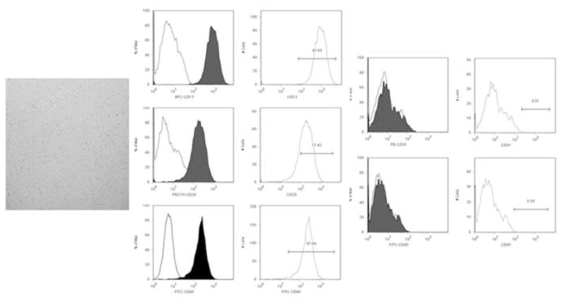
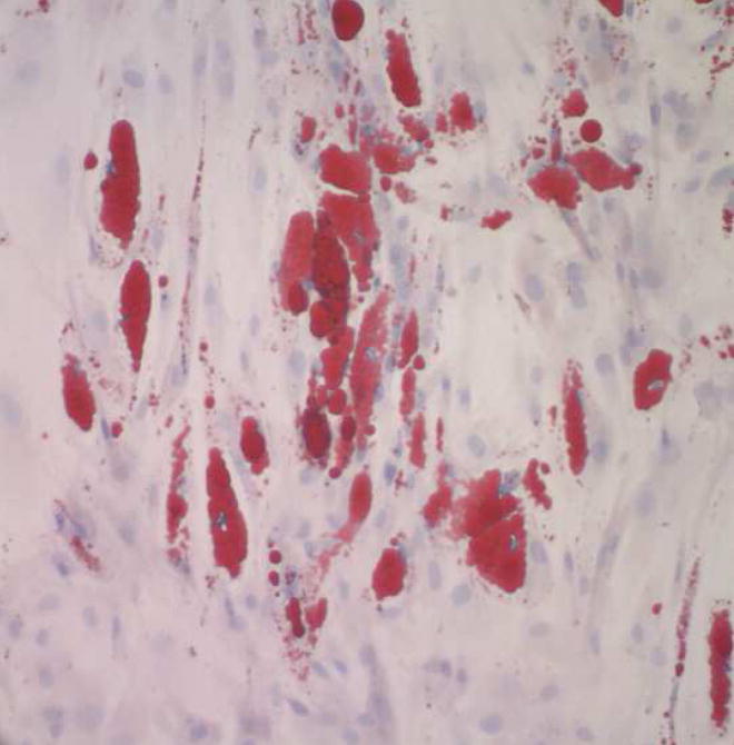
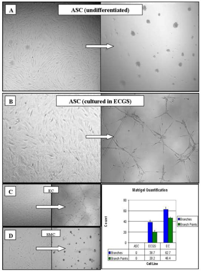
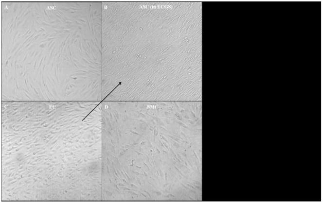
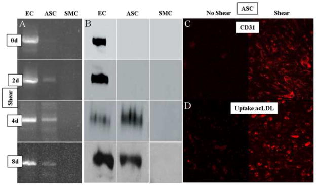
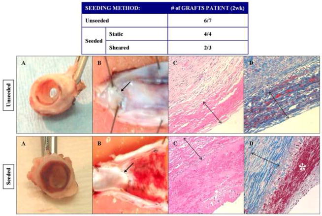
Materials and methods: Human ASC (CD13(+)29(+)90(+)31(-)45(-)) were isolated from periumbilical fat, cultured in ECGS media (for up to 3 wk), and exposed to physiological shear force (12 dynes for up to 8 d) in vitro. Endothelial phenotype was defined by cord formation on Matrigel, acetylated-low density lipoprotein (acLDL) uptake, and expression of nitric oxide synthase (eNOS), von Willebrand factor (vWF), and CD31 (platelet endothelial cell adhesion molecule, PECAM). Additionally, cell thrombogenicity was evaluated by seeding canine autologous ASC onto vascular grafts implanted within the canine arterial circulation for 2 wk.

Results: We found that undifferentiated ASC did not display any of the noted endothelial characteristics. After culture in ECGS, ASC formed cords in Matrigel but failed to take up acLDL or express the molecular markers. Subsequent exposure to shear resulted in stem cell realignment, acLDL uptake, and expression of CD31; eNOS and vWF expression was still not observed. Grafts seeded with cells grown in ECGS (+/- shear) remained patent (six of seven) at 2 wk but had a thin coat of fibrin along the luminal surfaces.

Conclusions: This study suggests that (1) ECGS and shear promote the expression of several endothelial characteristics in human adipose-derived stem cells, but not eNOS or vWF; (2) their combined effects appear synergistic; and (3) stem cells differentiated in ECGS appear mildly thrombogenic in vitro, possibly related, in part, to insufficient eNOS expression. Thus, while the acquisition of several endothelial characteristics by adult stem cells derived from adipose tissue suggests these cells are a viable source of autologous cells for cardiovascular regeneration, further stimulation/modifications are necessary prior to using them as a true endothelial cell replacement.


Figures

Figure 1
Figure 1. FACS analysis of human adipose-derived stem cells
Photomicrograph of stem cells immediately after isolation, culture and negative selection for CD31 and 45 (phase-contrast, 40x) demonstrating homogeneous, spindle-shaped morphology. Representative FACS analysis of an isolate used in this study illustrates expression of the mesenchymal stem cells markers CD13, 29 and 90. Negative selection successfully removed co-isolated CD31 (endothelial) and CD45 (mononuclear) cells, allowing for the conclusion that subsequent expression of endothelial characteristics were due to stem cell differentiation and not culture selection.
Figure 2
Figure 2. Differentiation of human adipose-derived stem cells into adipocytes
Photomicrograph of stem cells grown for 3.5 weeks (passage 3) in media promoting adipogenic differentiation (Oil Red O with Modified Mayer’s hematoxylin counter stain; 200x) reveals uptake of lipid within the cells. This finding demonstrates that cells used in this study are capable of differentiating into a cell line different than endothelial cells.
Figure 3
Figure 3. Effect of ECGS on the differentiation of human adipose-derived stem cells (ASC)
Photomicrographs of cells plated onto Matrigel (left panels) and subsequently cultured for 24 h (right panels; phase-contrast, 100x). Undifferentiated stem cells (A), naïve to ECGS, recoil and ball up in response to Matrigel, similar to smooth muscle cell (SMC) controls (D). In contrast, stem cells grown in ECGS for a minimum of 7 days (B) form cord-like structures similar to the endothelial cell (EC) controls (C).
Figure 4
Figure 4. Morphological effects of shear force on the differentiation of human adipose-derived stem cells (ASC)
Photomicrographs of cells exposed to 12 dynes of shear in vitro for 4 days (phase-contrast, 100x). The arrow indicates the direction of shear in all four panels. Undifferentiated stem cells (A), naïve to ECGS, remain randomly oriented, similar to smooth muscle cell (SMC) controls (D). In contrast, stem cells grown in ECGS for 2 weeks and subsequently exposed to shear (B) orient in the direction of flow similar to the endothelial cell (EC) controls (C).
Figure 5
Figure 5. Molecular effects of shear force on the differentiation of human adipose-derived stem cells
Panel A. RT-PCR results (CD31). In these experiments, endothelial (EC), differentiated stem (ASC), and smooth muscle (SMC) cells were exposed to shear for up to 8 days. Examination of the ASC results reveals the expression of CD31 message beginning 2 days after shear was introduced, and continuing throughout 8 days. Panel B. Immunoblot results (CD31). In experiments parallel to those in panel A, each of the cell lines were exposed to shear for up to 8 days. Examination of the ASC results reveals the expression of CD31 protein beginning 4 days after shear was introduced, and continuing throughout 8 days. Panel C. Immunohistochemical results (CD31). Fluorescent photomicrograph of differentiated ASC stained for CD31 before (left) and after 4 days (right) of shear demonstrating significant expression of this protein in response to shear (100x). Panel D. acLDL uptake. Fluorescent photomicrograph of differentiated ASC stained for acLDL before (left) and after 8 days (right) of shear demonstrating significant uptake in response to shear (100x).
Figure 6
Figure 6. In vivo evaluation of the thrombogenicity of human adipose-derived stem cells
In these experiments, autologous canine adipose-derived stem cells were cultured in ECGS and seeded onto canine decellularized vein grafts with (static) and without (shear) flow conditioning. Unseeded grafts were used as controls. The grafts were then implanted into the carotid circulation of the animal from which the stem cells were harvested. The Table reveals that patency of the grafts were similar amongst the seeded (6 of 7) and unseeded (6 of 7) groups. After explant, gross examination (Panels A and B) of the seeded grafts revealed a thin layer of thrombus throughout the graft, not seen on the unseeded grafts. The proximal anastomotic suture lines are marked with an arrow. Microscopic evaluation (Panels C and D, H&E and trichrome stain, respectively; 100x) confirms that this layer contains fibrin (red staining marked by * in Panel D), and is absent on the unseeded controls. The decellularized vein allografts (marked by double arrows in Panels C and D) are noted to be largely composed of collagen (blue staining in Panels D).

References

    1. Fukuda K, Yuasa S. Stem Cells as a Source of Regenerative Cardiomyocytes. Circ Res. 2006;98:1002–1013. - PubMed
    1. Wollert KC, Drexler H. Clinical Applications of Stem Cells for the Heart. Circ Res. 2005;96:151–163. - PubMed
    1. Urbich C, Aicher A, Heeschen C, Dernbach E, Hofmann WK, Zeiher AM, Dimmeler S. Soluble factors released by endothelial progenitor cells promote migration of endothelial cells and cardiac resident progenitor cells. Journal of Molecular and Cellular Cardiology. 2005;39:733–742. - PubMed
    1. Iwase T, Nagaya N, Fujii T, Itoh T, Murakami S, Matsumoto T, Kangawa K, Kitamura S. Comparison of angiogenic potency between mesenchymal stem cells and mononuclear cells in a rat model of hindlimb ischemia. Cardiovascular Research. 2005;66:543–551. - PubMed
    1. Campbell GR, Campbell JH. Development of tissue engineered vascular grafts. Curr Pharm Biotechnol. 2007;8:43–50. - PubMed

Publication types

MeSH terms