. 2009 Nov 4;97(9):2585-94.
doi: 10.1016/j.bpj.2009.08.028.

Human serum albumin inhibits Abeta fibrillization through a "monomer-competitor" mechanism

Affiliations

Human serum albumin inhibits Abeta fibrillization through a "monomer-competitor" mechanism

Julijana Milojevic et al. Biophys J. .

Abstract

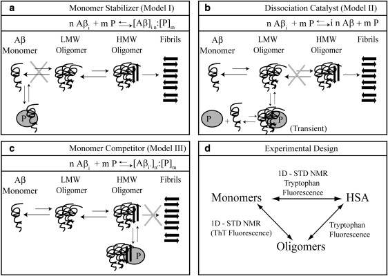
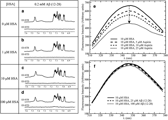
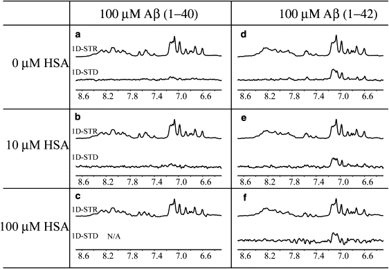
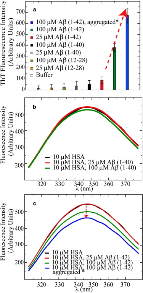
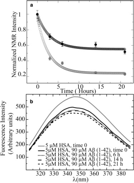
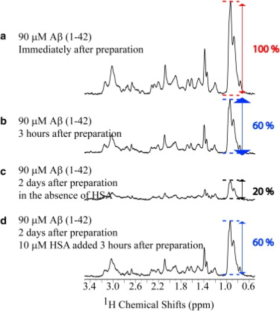
Human serum albumin (HSA) is not only a fatty acid and drug carrier protein, it is also a potent inhibitor of Abeta self-association in plasma. However, the mechanism underlying the inhibition of Abeta fibrillization by HSA is still not fully understood. We therefore investigated the Abeta-HSA system using a combined experimental strategy based on saturation transfer difference (STD) NMR and intrinsic albumin fluorescence experiments on three Abeta peptides with different aggregation propensities (i.e., Abeta(12-28), Abeta(1-40), and Abeta(1-42)). Our data consistently show that albumin selectively binds to cross-beta-structured Abeta oligomers as opposed to Abeta monomers. The HSA/Abeta oligomer complexes have K(D) values in the micromolar to submicromolar range and compete with the further addition of Abeta monomers to the Abeta assemblies, thus inhibiting fibril growth ("monomer competitor" model). Other putative mechanisms, according to which albumin acts as a "monomer stabilizer" or a "dissociation catalyst", are not supported by our data, thus resolving previous discrepancies in the literature regarding Abeta-HSA interactions. In addition, the model and the experimental approaches proposed here are anticipated to have broad relevance for the characterization of other systems that involve amyloidogenic peptides and oligomerization inhibitors.


Figures

Figure 1
Figure 1
Panels a–c depict possible hypothetical models for the mechanism of oligomerization inhibition by a generic inhibitory protein P. Aβ denotes the Aβ peptide in its monomeric state, whereas Aβi and Aβi' indicate Aβ oligomers. LMW and HMW Aβ refer to low- and high-MW Aβ oligomers, respectively. The letters n, m, and n′ refer to integer numbers that define the stoichiometry of the noncovalent complexes involving the Aβ peptide and the P protein. In both models I and II, oligomers are disrupted (i.e., “cleared”) by P. Whereas an Aβ oligomer-HSA complex in model II forms only transiently, in model III it does not clear the oligomers and HSA binds stably to them, preventing their further growth into larger assemblies. To include the possibility that the inhibitory binding protein partially converts large oligomers into a higher number of smaller oligomers, the subscripts i and n were replaced by i′ and n′ for the P-bound oligomers in model III. In any case, such oligomers must remain larger than the critical size required to interact with the inhibitory protein (denoted as ics in panel c), i.e., i n = in′ and ics < i′ < i. The cartoon representation of the models was used for clarity, but it does not imply a specific pathway for fibril formation or assign specific structures or stoichiometries for Aβ in different oligomerization or HSA-bound states. Panel d summarizes the experimental design to test models I–III. The STD NMR experiments mainly probe interactions with the low-MW components of the system (i.e., Aβ monomers); however, tryptophan fluorescence can probe HSA interactions with both Aβ monomers and oligomers. Due to HSA-ThT interactions, ThT fluorescence can be used to reliably probe cross-β-structured oligomers only in the absence of HSA.
Figure 2
Figure 2
(a–d) Effects of HSA on the STR and STD spectra of 0.2 mM Aβ(12–28) in 50 mM acetic acid-d4, pH 4.7, 10% D2O. The STR and STD spectra of 10 and 100 μM HSA solutions were subtracted from the protein peptide mixture spectra to remove residual HSA signal. All spectra were acquired at 700 MHz using a TCI CryoProbe and at 20°C. A 30-ms-long SL was used to minimize the residual HSA signal. All spectra were processed using a line-broadening factor of 3 Hz. Panels e and f depict the effects of aspirin and Aβ(12–28), respectively, on the emission intrinsic fluorescence spectra of HSA.
Figure 3
Figure 3
Effect of HSA on the STR and STD spectra of Aβ(1–40) and Aβ(1–42) samples. All peptide solutions were prepared at a 0.1 mM concentration in 20 mM potassium phosphate buffer, pH 7.4, 10% D2O. A 30-ms-long SL was used to minimize the HSA signal. The STR and STD spectra of the 10 and 100 μM HSA solutions were collected and then subtracted from the protein peptide mixture spectra. Although this subtraction was possible for the 100 μM Aβ(1–42) sample, it was not viable for the Aβ(1–40) sample due to the negligible STD effect arising from this peptide as compared to that originating from albumin. All spectra were acquired at 700 MHz using a TCI CryoProbe and at 20°C. The STR and STD spectra were processed using a line-broadening factor of 10 Hz. In panel f, at a 100 μM albumin concentration, the 30-ms SL becomes less effective at completely removing the protein signal, resulting in residual difference artifacts at a range of 7.4–7.8 ppm.
Figure 4
Figure 4
(a) Probing Aβ assemblies using ThT fluorescence. All samples were freshly prepared, with the exception of one 100 μM Aβ(1–42) sample (blue bar) that was aggregated for 3 h at 37°C before the ThT fluorescence measurements. This sample is denoted by an asterisk (). ThT was present in all samples at a 20 μM concentration. At least four measurements were collected for each sample, and the average values are reported. The error was calculated as the standard deviation of all measurements. (b) Interactions of HSA with Aβ(1–40) as probed by tryptophan fluorescence. (c) Interaction of HSA with Aβ(1–42) probed by HSA tryptophan fluorescence quenching at increasing Aβ(1–42) concentrations.
Figure 5
Figure 5
Time-dependent aggregation of 90 μM Aβ(1–42) in the absence (gray curve) and presence (black curve) of 5 μM HSA as monitored by NMR 1D NMR spectra with a 30-ms SL filter (a) and by intrinsic HSA tryptophan fluorescence (b). The experimental data were fitted using the offset decaying exponential: a × e-bt + c, where t is in hours and the ac parameters were obtained through nonlinear curve-fitting. The actual experimental data are plotted in solid circles, and the fitted values are shown in open circles. Between the readings, samples were incubated in a water bath at 37°C. The NMR intensities reported in panel a are normalized intensities of the methyl spectral region (0.6–1.1 ppm) measured as a function of time. The error was estimated from the spectral noise to be ∼5%. NMR experiments were acquired at 700 MHz at 37°C in 20 mM potassium phosphate, pH 7.4, 10% D2O, 0.02% NaN3.
Figure 6
Figure 6
Effect of a delayed addition of HSA on the aggregation profile of Aβ(1–42). Panels a and b show the 1D NMR spectra of 90 μM Aβ(1–42) immediately after preparation and after 3 h, respectively. In the absence of HSA, 2 days after the sample was prepared, ∼80% of the initial NMR signal is lost, as shown in panel c. However, when 10 μM of HSA were added 3 h after sample preparation, no NMR signal losses were observed even after 2 days (d). These spectra were recorded at 600 MHz and 37°C. In between acquisition sessions, samples were stored in a water bath at 37°C. Note that the Aβ(1–42) samples used here and in Fig. 5 came from different stock solutions.

References

    1. Cummings J.L. Alzheimer's disease. N. Engl. J. Med. 2004;351:56–67. - PubMed
    1. Haass C., Selkoe D.J. Cellular processing of β- amyloid precursor protein and the genesis of amyloid β -peptide. Cell. 1993;73:1039–1042. - PubMed
    1. Walsh D.M., Klyubin I., Fadeeva J.V., Cullen W.K., Anwyl R. Naturally secreted oligomers of the Alzheimer amyloid β-protein potently inhibit hippocampal long-term potentiation in vivo. Nature. 2002;416:535–539. - PubMed
    1. Dahlgren K.N., Manelli A.M., Stine W.K., Baker L.K., Jr., Krafft G.A. Oligomeric and fibrillar species of amyloid-β peptides differentially affect neuronal viability. J. Biol. Chem. 2002;277:32046–32053. - PubMed
    1. Zlokovic B.V. Clearing amyloid through the blood–brain barrier. J. Neurochem. 2004;89:807–811. - PubMed

Publication types