. 2009 Dec 15;587(Pt 24):5907-23.
doi: 10.1113/jphysiol.2009.179887.

Both synaptic and intrinsic mechanisms underlie the different properties of population bursts in the hippocampal CA3 area of immature versus adult rats

Affiliations

Both synaptic and intrinsic mechanisms underlie the different properties of population bursts in the hippocampal CA3 area of immature versus adult rats

Li-Rong Shao et al. J Physiol. .

Abstract

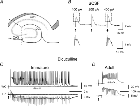
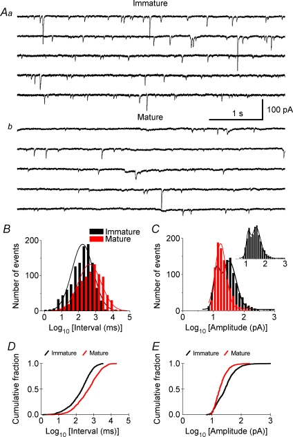
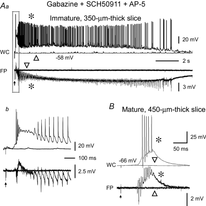
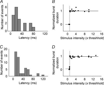
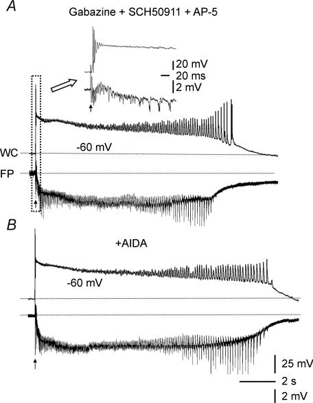
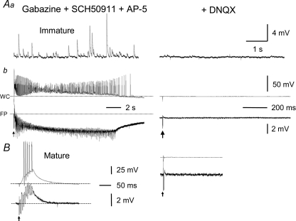
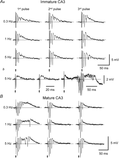
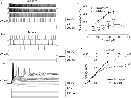
Pharmacological blockade of GABA(A) receptors on CA3 pyramidal cells in hippocampal slices from immature rats (i.e. second to third postnatal weeks), compared to CA3 slices from adult rats, is known to cause prolonged burst discharges (i.e. several seconds vs. tens of milliseconds). Synaptic and intrinsic mechanisms responsible for this developmental difference in burst duration were analysed in isolated minislices of the CA3 area. The frequency and amplitude of spontaneous EPSCs in CA3 pyramidal cells were greater in slices from immature than mature rats. In the presence of GABA(A)- and GABA(B)-receptor antagonists, the burst discharges of immature CA3 pyramidal cells were still prolonged in thinner slices (350 microm, vs. 450 microm in adults, to compensate for developmental differences in neuronal density) and in NMDA- and mGlu1-receptor antagonists. The AMPA receptor antagonist DNQX blocked the remaining burst discharges, suggesting that differences in recurrent excitatory circuits contributed to the prolonged bursts of immature CA3 pyramidal cells. In slices from immature versus adult rats, the CA3 recurrent synaptic responses showed potentiation to repetitive stimulation, suggestive of a lower transmitter release probability. The intrinsic firing ability was greater in CA3 pyramidal neurons from immature than adult rats, and the medium-duration afterhyperpolarization was smaller. These data suggest that, compared to adults, the CA3 area of immature rats contains a more robust recurrent excitatory synaptic network, greater intrinsic membrane excitability, and an increased capacity for sustained transmitter release, which together may account for the more prolonged network bursts in immature versus adult CA3.


Figures

Figure 1
Figure 1. Prolonged population bursts after bicuculline treatment in CA3 minislices of immature versus adult rats
A, diagram showing CA3 minislices used in this study. Dashed lines indicate knife cuts to isolate the CA3 area from CA1 and dentate gyrus. B shows that in normal aCSF, synaptic stimulation in CA3 minislices from immature rats only elicited a field EPSP and a single population spike and never evoked epileptiform bursts, regardless of the stimulus intensity (the three responses were evoked at 100 μA, 200 μA and 400 μA). Arrows indicate stimulations. Boxed parts in B are shown in expanded scales below. Note the difference in the amplitude of population spikes at different stimulus intensities (i.e. 100 μA and 400 μA). C, simultaneous whole-cell (WC, current-clamp mode) and field-potential (FP) recordings showing evoked population bursts in bicuculline (30 μm) in CA3 minislices from immature rats. These bursts typically contained an initial depolarization followed by rhythmic afterdischarges lasting for several seconds. D, by contrast, the evoked population bursts that occurred in CA3 slices of mature rats were composed of a single burst that lasted for a few hundred milliseconds.
Figure 2
Figure 2. Spontaneous excitatory postsynaptic currents (sEPSCs) in CA3 pyramidal neurons from immature and mature rats
A, representative sEPSCs recorded in CA3 neurons from immature (14 d, Aa) and adult (93 d, Ab) rats. Five consecutive traces (i.e. 5 s each) from continuous recordings of sEPSCs are shown in each panel. The sEPSCs in slices from immature rat (Aa) were more frequent and of larger amplitude than the sEPSCs recorded in adult slices (Ab). B, combined histograms showing the distribution of sEPSC intervals (milliseconds in log value, bin = 0.2) for the immature (black) and adult (red) groups. More sEPSCs in the immature group had short inter-event intervals (i.e. left side of histogram), and the peak of the histogram was shifted to the left (i.e. shorter interval) compared to that in the adult group. C, histograms showing the logarithmic distribution of sEPSC amplitudes for the immature (black) and adult (red) groups. The histogram of the immature group appeared to fit a two-peak distribution (inset), and the second peak (i.e. larger amplitude sEPSCs) was largely absent in the adult group. D and E, Kolmogorov–Smirnov test showing the cumulative probability of sEPSC intervals (D) and sEPSC amplitudes (E) was significantly different between immature (black) and mature (red) groups (P < 0.001). Note that 100 sEPSCs from each cell were pooled for constructing histograms and for calculating cumulative probability, except for a few cells that had fewer than (but close to) 100 sEPSCs. The peaks of histograms were fitted with a Gaussian distribution.
Figure 3
Figure 3. Prolonged population bursts were present in CA3 minislices from immature rats after pharmacological blockade of GABAB and NMDA receptors
Simultaneous whole-cell (WC, upper traces) and field-potential (FP, lower traces) recordings showing the response of a 350 μm-thick CA3 minislice from a 10-day-old rat (Aa) and a 450 μm-thick CA3 minislice from a 99-day-old rat (B) to synaptic stimulation in the presence of a mixture of antagonists of GABAA (gabazine, 10 μm), GABAB (SCH50911, 10 μm) and NMDA (AP-5, 50 μm) receptors. The immature CA3 slice in Aa responded to synaptic stimulation at threshold intensity (arrow) with a 15 s long population burst with rhythmic afterdischarges. The initial section of the burst (i.e. boxed segment) is shown at an expanded time scale in Ab. The responses were all-or-none, such that stimuli of the same intensity evoked either an entire prolonged burst (asterisks) or no burst (triangles). Note the long latency from stimulation (arrow) to the onset of the burst in Ab (∼75 ms). In contrast, in CA3 slices from mature rats, synaptic stimulation at threshold intensity (arrow) evoked brief population bursts (B). Note the all-or-none property of the response and the long latency (∼53 ms) at threshold stimulus intensity (20 μA).
Figure 4
Figure 4. The long-and-variable latency and all-or-none characteristics of the CA3 population bursts from immature and mature rats
A, histogram showing the distribution of the latency values of 18 bursts pooled from 8 CA3 minislices from immature rats. The latency values ranged from 5 to 75 ms, suggesting these bursts were multi-synaptic responses. Bin = 10 ms. B, pooled data from 9 CA3 minislices from immature rats showing the all-or-none nature of the prolonged network bursts; that is, stimulation below or at threshold intensity (≤ 1) evoked no bursts (duration = 0), while threshold or slight supra-threshold stimulations (≥1) elicited full-length bursts (duration = 1), and a further increase of stimulus intensity up to 10 times threshold no longer significantly increased burst duration. The vertical dashed line indicates the threshold (i.e. 1) and the short and long horizontal lines indicate the lack of a burst (0) and the emergence of a full length burst (1) in response to threshold stimulation, respectively. C, histogram showing the distribution of the latency values from 51 bursts pooled from 9 slices from adult rats, which ranged from 11 to 131 ms, also suggesting the poly-synaptic nature of the brief epileptiform bursts. Bin = 10 ms. D, data pooled from 9 slices illustrating that the brief population bursts in adult CA3 minislices were also all-or-none.
Figure 5
Figure 5. The prolonged epileptiform bursts of immature CA3 pyramidal cells were also present in the mGlu1-receptor antagonist, AIDA
A, whole-cell (WC, upper trace) and field-potential (FP, lower trace) recordings showing a prolonged evoked burst in a CA3 minislice from an 11-day-old rat in gabazine, SCH50911 and AP-5. Inset at the top shows the boxed part of this burst at an expanded time scale. B, bath-application of the selective mGlu1 receptor antagonist AIDA (500 μm) did not reduce burst duration in this slice.
Figure 6
Figure 6. All sEPSPs and epileptiform bursts were completely abolished by the AMPA/kainate-receptor antagonist, DNQX
A, frequent sEPSPs as large as 10 mV (Aa, left panel) were present and prolonged epileptiform bursts (Ab, left panel) were consistently elicited in an immature CA3 minislice bathed with a mixture of gabazine (10 μm), SCH50911 (10 μm) and AP-5 (50 μm). DNQX (50 μm) completely abolished the sEPSPs and prolonged population bursts (Aa and Ab, right panels), confirming that the prolonged bursts under these conditions (i.e. prior blockade of GABAA, GABAB and NMDA receptors) were solely mediated by AMPA/kainate receptors. B, DNQX similarly eliminated the brief bursts evoked in mature CA3 minislices. Arrows indicate stimulations. Dotted lines in A and B indicate baseline potentials.
Figure 7
Figure 7. Responsiveness of the CA3 recurrent synapses to repetitive stimulation in slices from immature and mature rats
CA3 recurrent synapses were antidromically activated in the presence of gabazine (10 μm), SCH50911 (10 μm), AP-5 (μm) and elevated [Mg2+]o (4 mm). Repetitive stimulations at different intervals (0.3, 1 and 5 Hz) were used to test the dynamic properties of transmitter release. Aa, field-potential recordings showed that the initial responses were comparatively small in immature CA3 slices, but they tended to increase in later responses, particularly at short intervals (i.e. 5 Hz). Ab, some slices responded to the initial stimulation with only a small field EPSP, but developed larger responses with multiple population spikes and longer duration during later stimulation. B, in contrast, in mature CA3 slices, the initial responses were on average larger than those in immature slices, and they tended to decrease during repetitive activation.
Figure 8
Figure 8. Intrinsic firing in CA3 pyramidal neurons from immature and adult rats
A, whole-cell recordings showing that CA3 pyramidal neurons from immature rats were able to fire action potentials over the entire duration of a depolarizing current pulse (i.e. 15 s) with little adaptation. B, in contrast, CA3 pyramidal neurons from mature rats responded to the same or larger current injections with fewer action potentials (Ba), or only generated action potentials during the first few hundred milliseconds of the current pulses (Bb). A strong rectification to depolarizing current pulses was seen in the mature CA3 neurons (Bb), which was not present in immature CA3 pyramidal cells. C, summary plot showing the difference in the input–output relations in CA3 pyramidal neurons from immature and mature rats. *P < 0.05; **P < 0.01; ***P < 0.001, Student's two-tailed unpaired t test. D, summary plot showing that the immature CA3 pyramidal neurons displayed a nearly ohmic current–voltage response, whereas adult CA3 pyramidal neurons showed a clear rectification to depolarizing current injections (i.e. deviated from the hypothetical linear response as the dotted line indicates), which limited their ability to fire repetitively. All experiments were performed in the presence of gabazine (10 μm), SCH50911 (10 μm), AP-5 (50 μm) and DNQX (50 μm), which blocked GABAergic and glutamatergic synaptic transmission.
Figure 9
Figure 9. Afterhyperpolarizations (AHPs) in CA3 neurons from immature and mature rats
A, most of the immature CA3 neurons displayed little or no discernable AHP after a burst of 4–5 action potentials (upper traces) induced by an 80 ms triangular depolarizing current injection (bottom trace). B, some of the mature CA3 neurons showed a medium-duration AHP (mAHP) and negligible slow AHP (sAHP). The membrane potential of the neurons was held at depolarized levels (−50 mV to −40 mV) to increase the driving force for AHP (K+ conductance). C, summary data showing the amplitude of mAHP and sAHP in the immature and mature groups (**P < 0.01, Student's two-tailed unpaired t test).

Comment in

References

    1. Belleau ML, Warren RA. Postnatal development of electrophysiological properties of nucleus accumbens neurons. J Neurophysiol. 2000;84:2204–2216. - PubMed
    1. Ben-Ari Y, Cherubini E, Krnjevic K. Changes in voltage dependence of NMDA currents during development. Neurosci Lett. 1988;94:88–92. - PubMed
    1. Ben-Ari Y, Khazipov R, Leinekugel X, Caillard O, Gaiarsa JL. GABAA, NMDA and AMPA receptors: a developmentally regulated ‘menage a trois’. Trends Neurosci. 1997;20:523–529. - PubMed
    1. Ben-Ari Y. Excitatory actions of GABA during development: the nature of the nurture. Nat Rev Neurosci. 2002;3:728–739. - PubMed
    1. Brumback AC, Staley KJ. Thermodynamic regulation of NKCC1-mediated Cl– cotransport underlies plasticity of GABAA signalling in neonatal neurons. J Neurosci. 2008;28:1301–1312. - PMC - PubMed

Publication types