. 2009 Nov 17;106(46):19599-604.
doi: 10.1073/pnas.0907935106. Epub 2009 Nov 2.

HDAC6 is a target for protection and regeneration following injury in the nervous system

Affiliations

HDAC6 is a target for protection and regeneration following injury in the nervous system

Mark A Rivieccio et al. Proc Natl Acad Sci U S A. .

Abstract

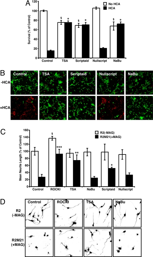
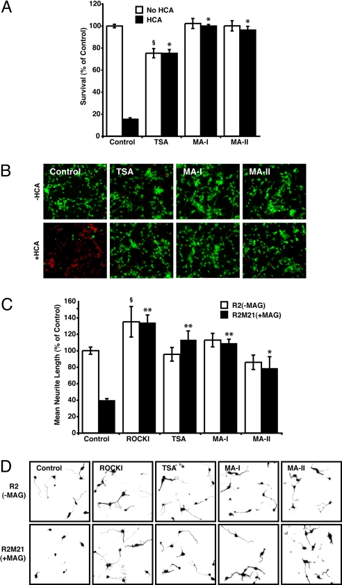
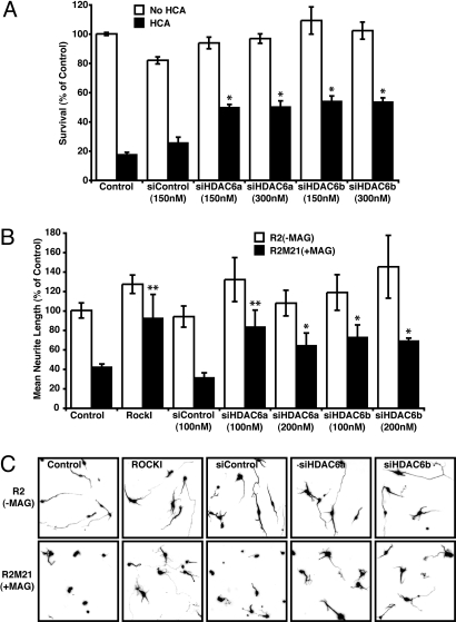
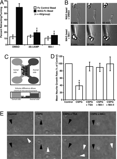
Central nervous system (CNS) trauma can result in tissue disruption, neuronal and axonal degeneration, and neurological dysfunction. The limited spontaneous CNS repair in adulthood and aging is often insufficient to overcome disability. Several investigations have demonstrated that targeting HDAC activity can protect neurons and glia and improve outcomes in CNS injury and disease models. However, the enthusiasm for pan-HDAC inhibition in treating neurological conditions is tempered by their toxicity toward a host of CNS cell types -a biological extension of their anticancer properties. Identification of the HDAC isoform, or isoforms, that specifically mediate the beneficial effects of pan-HDAC inhibition could overcome this concern. Here, we show that pan-HDAC inhibition not only promotes neuronal protection against oxidative stress, a common mediator of injury in many neurological conditions, but also promotes neurite growth on myelin-associated glycoprotein and chondroitin sulfate proteoglycan substrates. Real-time PCR revealed a robust and selective increase in HDAC6 expression due to injury in neurons. Accordingly, we have used pharmacological and genetic approaches to demonstrate that inhibition of HDAC6 can promote survival and regeneration of neurons. Consistent with a cytoplasmic localization, the biological effects of HDAC6 inhibition appear transcription-independent. Notably, we find that selective inhibition of HDAC6 avoids cell death associated with pan-HDAC inhibition. Together, these findings define HDAC6 as a potential nontoxic therapeutic target for ameliorating CNS injury characterized by oxidative stress-induced neurodegeneration and insufficient axonal regeneration.


Conflict of interest statement

The authors declare no conflict of interest.

Figures

Fig. 1.
Fig. 1.
Pharmacological HDAC inhibition protects primary neurons from oxidative stress and promotes neurite outgrowth in the presence of MAG. (A) Neuronal viability after treatment with homocysteic acid (HCA; 5 mM) with or without pan-HDAC inhibitors TSA (0.66 μM), Scriptaid (6.13 μM), Nullscript (6.13 μM), and sodium butyrate (NaBu; 1 mM) for 24 h. *, Significant protection compared to HCA treatment alone P < 0.001. §, Significant death compared to non-HCA control P < 0.001. (B) Live/Dead staining of treated rat primary cortical cultures. Live cells are identified by green fluorescence, whereas dead cells identified by red fluorescence. (C and D) Mean neurite length measured after 24 h coculture with either R2 or R2M21+MAG CHO cells in the presence or absence of the RhoA kinase Inhibitor (ROCKI), Y27632 (10 μM), or the HDAC inhibitors, TSA, NaBu, Scriptaid, or Nullscript. Neurites were identified by neuron-specific β-III tubulin immunostaining and fluorescence microscopy (D), and measured by using Metamorph (C). Significant increase in neurite length relative to nontreated R2M21+MAG denoted by *, P < 0.05, **, P < 0.01, or ***, P < 0.001. Significant increase in neurite length relative to nontreated R2 denoted by §, P < 0.05.
Fig. 2.
Fig. 2.
Pharmacological inhibitors selective for HDAC6 are protective in oxidatively stressed neurons, and promote neurite outgrowth in the presence of MAG. (A) Neuronal viability after treatment with homocysteic acid (HCA; 5 mM) with or without the pan-HDAC inhibitor TSA (0.66 μM), or the HDAC6-selective inhibitors MA-I (10 μM), and MA-II (10 μM) for 24 h. Viability was measured by using MTT assay. *, Significant protection compared to HCA treatment alone P < 0.001. §, Significant death compared to non-HCA control P < 0.001. (B) Live/Dead staining of treated rat primary cortical cultures. (C and D) Mean neurite length of cortical neurons measured after 24 h coculture with either R2 and R2M21+MAG cells in the presence or absence of the RhoA kinase Inhibitor (ROCKI), Y27632 (10 μM), the pan-HDAC inhibitor, TSA, or the HDAC6-selective inhibitors MA-I, and MA-II. Neurites were identified by neuron-specific β-III tubulin immunostaining and fluorescence microscopy (D), and measured by using Metamorph (C). Significant increase in neurite length relative to nontreated R2M21+MAG denoted by *, P < 0.05 or **, P < 0.001. Significant increase in neurite length relative to nontreated R2 denoted by §, P < 0.05.
Fig. 3.
Fig. 3.
Targeted knockdown of HDAC6 using RNAi is protective and promotes neurite outgrowth. (A) Rat primary cortical neuron viability after treatment with homocysteic acid (HCA; 5 mM) with or without a nonspecific 21-nt duplex (siControl), or HDAC6 specific siRNAs (siHDAC6a and siHDAC6b) for 24 h. Viability was measured by using MTT assay. *, Significant protection compared to HCA treatment alone P < 0.001. (B and C) Mean neurite length of cortical neurons measured 24 h after coculture with either R2 and R2M21+MAG cells in the presence or absence of the RhoA kinase Inhibitor (ROCKI), Y27632 (10 μM), or nonspecific 21-nt duplex (siControl), or HDAC6 specific siRNAs (siHDAC6a and siHDAC6b). Neurites were identified by neuron-specific β-III tubulin immunostaining and fluorescence microscopy (C), and measured by using Metamorph (B). Significant increase in neurite length relative to nontreated R2M21+MAG denoted by *, P < 0.05 or **, P < 0.001.
Fig. 4.
Fig. 4.
Pharmacological inhibitors selective for HDAC6 promote dorsal root ganglion neuron axonal outgrowth in the presence of MAG and CSPGs. (A) Percent axonal retraction of rat adult dorsal root ganglion neurons when presented to Fc control or MAG-Fc coated microspheres in the presence of vehicle control (DMSO), dibutyryl cAMP or the HDAC6-selective inhibitor, MA-I. *, Significant decrease in neurite retraction/turning relative to nontreated control P < 0.001. (B) Representative micrographs at 0, 30, and 60 min after the presentation of MAG to the growth cone of a DRG axon. (C) Schematic of microfluidic-based culture platform adapted from Taylor et al. (31). The culture chamber consists of somal and axonal compartments connected by microgrooves. Rat DRG neurons are added to the somal side and axonal growth is guided into the axonal side through the microgrooves. Volume difference between the somal side and axonal side allows chemical microenvironments to be isolated. (D) Growth rate of rat adult dorsal root ganglion axons exposed to laminin (Control) or CSPG/laminin in the presence of DMSO vehicle, TSA, or the HDAC6-selective inhibitors, MA-I and MA-II. Growth rates of axons exposed to CSPG/laminin in the presence HDAC inhibitors were not significantly different to growth rates of axons exposed to laminin-only. *, Significant decrease in neurite growth relative to laminin-only controls P < 0.05 by. (E) Representative micrographs of axons in the CSPG- containing axonal compartment, at 2 h (Top) and 5 h (Bottom) after treatment. Black arrows show axonal growth cones at 2 h. White arrows show axonal growth cones at 5 h.

References

    1. Anderson MF, Sims NR. The effects of focal ischemia and reperfusion on the glutathione content of mitochondria from rat brain subregions. J Neurochem. 2002;81:541–549. - PubMed
    1. Young W. Secondary injury mechanisms in acute spinal cord injury. J Emerg Med. 1993;11(Suppl 1):13–22. - PubMed
    1. Springer JE. Apoptotic cell death following traumatic injury to the central nervous system. J Biochem Mol Biol. 2002;35:94–105. - PubMed
    1. Blight AR. Cellular morphology of chronic spinal cord injury in the cat: Analysis of myelinated axons by line-sampling. Neuroscience. 1983;10:521–543. - PubMed
    1. Fehlings MG, Tator CH. The relationships among the severity of spinal cord injury, residual neurological function, axon counts, and counts of retrogradely labeled neurons after experimental spinal cord injury. Exp Neurol. 1995;132:220–228. - PubMed

Publication types

MeSH terms

LinkOut - more resources