. 2009 Dec;119(12):3713-22.
doi: 10.1172/JCI40053. Epub 2009 Nov 2.

Mfge8 diminishes the severity of tissue fibrosis in mice by binding and targeting collagen for uptake by macrophages

Affiliations

Mfge8 diminishes the severity of tissue fibrosis in mice by binding and targeting collagen for uptake by macrophages

Kamran Atabai et al. J Clin Invest. 2009 Dec.

Abstract

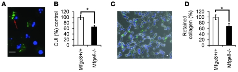
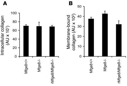
Milk fat globule epidermal growth factor 8 (Mfge8) is a soluble glycoprotein known to regulate inflammation and immunity by mediating apoptotic cell clearance. Since fibrosis can occur as a result of exaggerated apoptosis and inflammation, we set out to investigate the hypothesis that Mfge8 might negatively regulate tissue fibrosis. We report here that Mfge8 does decrease the severity of tissue fibrosis in a mouse model of pulmonary fibrosis; however, it does so not through effects on inflammation and apoptotic cell clearance, but by binding and targeting collagen for cellular uptake through its discoidin domains. Initial analysis revealed that Mfge8-/- mice exhibited enhanced pulmonary fibrosis after bleomycin-induced lung injury. However, they did not have increased inflammation or impaired apoptotic cell clearance after lung injury compared with Mfge8+/+ mice; rather, they had a defect in collagen turnover. Further experiments indicated that Mfge8 directly bound collagen and that Mfge8-/- macrophages exhibited defective collagen uptake that could be rescued by recombinant Mfge8 containing at least one discoidin domain. These data demonstrate a critical role for Mfge8 in decreasing the severity of murine tissue fibrosis by facilitating the removal of accumulated collagen.


Figures

Figure 1
Figure 1. Mfge8 expression is induced by lung injury.
(A and B) Lung sections taken from adult wild-type mice were stained with anti-Mfge8 antibody. Mfge8 was expressed in the alveolar interstitium (arrow in A) and pulmonary endothelium (arrow in B). Scale bars: 10 μm. (C and D) Alveolar macrophages were obtained by BAL after saline administration (C) or 5 days after bleomycin administration (5 U/kg) (D). Mfge8 staining was present in macrophages from saline-treated animals (C), and the intensity of expression increased after bleomycin treatment (D). Scale bars: 10 μm. (E) Ten micrograms of protein from total lung homogenates taken at the indicated days after bleomycin treatment (1.1 U/kg) or 7 days after saline treatment was loaded for a Western blot using an anti-Mfge8 antibody. An antibody against Crk was used to demonstrate equal loading of protein. (F) One microgram of protein from lung homogenates obtained from human patients with IPF was loaded for a Western blot using an anti-lactadherin antibody (Lact.). Control samples were from lungs rejected for transplantation. An antibody against Crk was used to demonstrate loading of protein.
Figure 2
Figure 2. Mfge8–/– mice develop exaggerated pulmonary fibrosis after injury.
(A and B) Picrosirius red staining of lung sections from Mfge8+/+ (A) and Mfge8–/– (B) mice taken 28 days after bleomycin administration (1.1 U/kg). Scale bars: 100 μm. (C and D) Biochemical analysis of bleomycin-induced pulmonary fibrosis. Pulmonary fibrosis measured by total lung hydroxyproline content at baseline and 28 days after bleomycin treatment in mice in a mixed-strain background (C) or pure-strain background (D). Data are presented as mean ± SEM (n = 6–8 for saline [Sal] and 10–14 for bleomycin [Bleo] treatment groups). *P = 0.004 (C) and *P = 0.01 (D) using Student’s t test to compare bleomycin-treated Mfge8+/+ and Mfge8–/– mice.
Figure 3
Figure 3. Mfge8–/– mice have intact apoptotic cell clearance in vivo after bleomycin treatment.
(A) Apoptotic thymocytes were instilled intratracheally and the phagocytic index of alveolar macrophages obtained by BAL 30 minutes after instillation was similar in Mfge8–/– and Mfge8+/– controls (n = 5–6). (B) Tissue sections taken from Mfge8–/– and Mfge8+/– mice at the indicated hours and days after bleomycin treatment (5 U/kg) were stained by TUNEL assay, and the number of apoptotic cells per ×200 fields (15 fields) was quantified (n = 4–10). (C) Sections taken from Mfge8–/– and Mfge8+/+ mice 9 days after bleomycin (1.1 U/kg) treatment were stained by TUNEL as described in B (n = 3–4). (DF) Cytospin preparations from BAL samples after bleomycin treatment (5 U/kg) were stained with Diff-Quick (D) or TUNEL (F), and the number of apoptotic cell ingestions (arrows in D; and E) and percentage of free apoptotic nuclei (F) were quantified (n = 5–8). There was no difference in number of TUNEL-positive cells, alveolar macrophage phagocytic index, or free apoptotic nuclei between Mfge8–/– and control samples. All comparisons were made using Student’s t test, and data are expressed as mean ± SEM.
Figure 4
Figure 4. Mfge8 mediates collagen uptake in vitro.
(A) Primary alveolar macrophages were cultured for 30 minutes with FITC-conjugated type I collagen, and uptake was evaluated by fluorescence microscopy (red arrows). Scale bar: 10 μm. (B) Ingestion of collagen was quantified as the collagen uptake index (CUI: number of macrophages with ingestions divided by the total number of macrophages counted). The addition of unlabeled type I collagen (25, 50, 150 μg/ml) inhibited uptake of FITC-conjugated collagen in a dose-dependent fashion (*P < 0.001, 1-way ANOVA with Bonferroni t test for multiple comparisons; n = 3–4; data are expressed as percent control relative to wild-type uptake). (C) Alveolar macrophages from Mfge8–/– mice had significantly impaired collagen uptake index as compared with Mfge8+/+ alveolar macrophages. (*P = 0.001, 1-way ANOVA with Bonferroni t test for multiple comparisons; n = 3–5). Addition of rMfge8 (μg/ml) rescued collagen uptake in Mfge8–/– alveolar macrophages (**P = 0.007, ***P = 0.023). (D) Addition of rMfge8 (μg/ml) increased collagen uptake in Mfge8+/+ alveolar macrophages under serum-starved conditions (*P = 0.009, Student’s t test to compare indicated columns; n = 5–6). Data are presented as mean ± SEM.
Figure 5
Figure 5. Mfge8 mediates collagen uptake in vivo.
(A) Alveolar macrophages obtained by BAL 30 minutes after intratracheal injection of FITC-conjugated type I collagen were examined by fluorescence microscopy for collagen uptake (red arrow). Scale bar: 10 μm. (B) Mfge8–/– alveolar macrophages had significantly impaired collagen uptake in vivo (*P = 0.009, Student’s t test; n = 6). (C) Frozen sections taken from lungs after intratracheal collagen injection were counterstained with DAPI, and the number of retained collagen particles divided by the total number of nuclei in each section was quantified. Scale bar: 10 μm. (D) Mfge8–/– lungs retained significantly fewer collagen particles (*P = 0.047 using a Student’s t test, n = 4). Data are presented as mean ± SEM.
Figure 6
Figure 6. Mfge8–/– fibroblasts do not have impaired collagen uptake.
(A and B) Primary lung fibroblasts from Mfge8–/– and Mfge8+/+ mice at passage 4 were incubated for 90 minutes with FITC-conjugated type l collagen, and unbound/uningested collagen was removed with multiple washes. Cells were then incubated with 50 μg/ml trypsin and 50 μg/ml proteinase K to remove membrane-bound collagen. Collagen in the membrane-bound portion (supernatant after enzymatic treatment) and intracellular portion (pellet remaining after enzymatic treatment) was quantified by a spectrofluorometer. There was no difference in membrane-bound (A) or intracellular (B) collagen with or without the addition of rMfge8 (13 μg/ml). Data are presented as mean ± SEM.
Figure 7
Figure 7. The first discoidin domain of Mfge8 is sufficient for collagen binding and uptake.
(A) Constructs containing full-length (Fl) Mfge8, Mfge8 lacking the terminal discoidin domain (Dd1), or both discoidin domains (Ndd) fused to a huFc domain were immobilized on a Biacore CM5 chip, and binding to increasing doses of collagen was evaluated. P/T represents a domain present in the long isoform of Mfge8 that is rich in proline and threonine. (B) Collagen bound full-length Mfge8 in a dose-dependent fashion with a Kd of 733 nM. (C) Flow plot demonstrating dose-dependent binding of collagen to immobilized full-length construct. Black line, 16 nM; filled squares, 31 nM; open diamonds, 63 nM; filled circles, 125 nM; open triangles, 250 nM; filled diamonds, 500 nM. (D) Flow plot demonstrating dose-dependent binding of collagen to immobilized Dd1 construct. (E) Flow plot demonstrating no binding of collagen to the Ndd construct. (F) The ability of constructs (13 μg/ml) to rescue the defect in alveolar macrophage collagen uptake was evaluated in vitro. Dd1 construct rescued Mfge8–/– alveolar macrophage collagen uptake, while the Ndd construct had no significant effect (*P = 0.01, 1-way ANOVA with Bonferroni t test; n = 3–4). Data are presented as mean ± SEM.

References

    1. Raghu G., Striker L.J., Hudson L.D., Striker G.E. Extracellular matrix in normal and fibrotic human lungs. Am. Rev. Respir. Dis. 1985;131:281–289. - PubMed
    1. Chua F., Gauldie J., Laurent G.J. Pulmonary fibrosis: searching for model answers. Am. J. Respir. Cell Mol. Biol. 2005;33:9–13. doi: 10.1165/rcmb.2005-0062TR. - DOI - PubMed
    1. Hammar S.P., Winterbauer R.H., Bockus D., Remington F., Friedman S. Idiopathic fibrosing alveolitis: a review with emphasis on ultrastructural and immunohistochemical features. Ultrastruct. Pathol. 1985;9:345–372. doi: 10.3109/01913128509074587. - DOI - PubMed
    1. Wynn T.A. Cellular and molecular mechanisms of fibrosis. J. Pathol. 2008;214:199–210. doi: 10.1002/path.2277. - DOI - PMC - PubMed
    1. Gross T.J., Hunninghake G.W. Idiopathic pulmonary fibrosis. N. Engl. J. Med. 2001;345:517–525. doi: 10.1056/NEJMra003200. - DOI - PubMed

Publication types

MeSH terms