. 2009 Dec;119(12):3703-12.
doi: 10.1172/JCI39845. Epub 2009 Nov 2.

Latent TGF-beta-binding protein 4 modifies muscular dystrophy in mice

Affiliations

Latent TGF-beta-binding protein 4 modifies muscular dystrophy in mice

Ahlke Heydemann et al. J Clin Invest. 2009 Dec.

Erratum in

  • J Clin Invest. 2010 Feb;120(2):645

Abstract

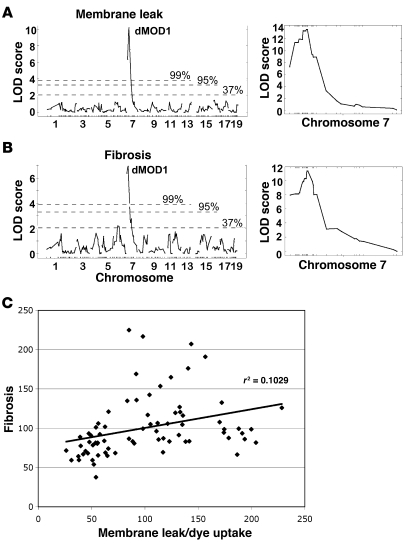
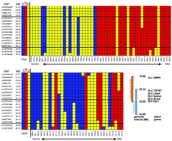
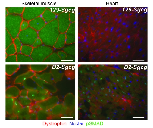
Most single-gene diseases, including muscular dystrophy, display a nonuniform phenotype. Phenotypic variability arises, in part, due to the presence of genetic modifiers that enhance or suppress the disease process. We employed an unbiased mapping approach to search for genes that modify muscular dystrophy in mice. In a genome-wide scan, we identified a single strong locus on chromosome 7 that influenced two pathological features of muscular dystrophy, muscle membrane permeability and muscle fibrosis. Within this genomic interval, an insertion/deletion polymorphism of 36 bp in the coding region of the latent TGF-beta-binding protein 4 gene (Ltbp4) was found. Ltbp4 encodes a latent TGF-beta-binding protein that sequesters TGF-beta and regulates its availability for binding to the TGF-beta receptor. Insertion of 12 amino acids into the proline-rich region of LTBP4 reduced proteolytic cleavage and was associated with reduced TGF-beta signaling, decreased fibrosis, and improved muscle pathology in a mouse model of muscular dystrophy. In contrast, a 12-amino-acid deletion in LTBP4 was associated with increased proteolysis, SMAD signaling, and fibrosis. These data identify Ltbp4 as a target gene to regulate TGF-beta signaling and modify outcomes in muscular dystrophy.


Figures

Figure 1
Figure 1. Mice lacking γ-sarcoglycan (Sgcg-null mice) serve as a model for muscular dystrophy.
The Sgcg-null allele displays a mild phenotype in the 129 background strain (129-Sgcg) and a severe phenotype in the D2 strain (D2-Sgcg). Sgcg mice from the two backgrounds were interbred (F2-Sgcg), and the F2-Sgcg animals displayed an intermediate phenotype. (A) Muscle membrane leak in the quadriceps muscle was measured by Evans blue dye uptake in the 2 parental strains (129-Sgcg and D2-Sgcg) and the intercrossed F2-Sgcg generation. (B) Quadriceps muscle fibrosis was measured by determining HOP content in the two parental strains; the F2-Sgcg mice also revealed an intermediate phenotype. (C) An example of the phenotypic range is shown from diaphragm muscles from 2 F2-Sgcg animals (the two left panels) and 1 wild-type control (right panel). Dye uptake (blue) and fibrotic replacement (white) are shown. (D) Grip strength was measured in 8-week-old animals of the 129 and D2 backgrounds with and without the Sgcg-null allele. The D2-Sgcg mice were weaker than 129-Sgcg mice, while the parental strains without the muscular dystrophy gene were not significantly different.
Figure 2
Figure 2. The pathological muscular dystrophy traits of membrane leak and fibrosis independently map to the dMOD1 locus on chromosome 7.
(A) lod scores are shown for 328 SNPs for the trait of membrane leak (dye uptake) in the quadriceps muscles. dMOD1 maps to chromosome 7 with a peak lod of 10.17. (B) lod scores across the genome correlated to fibrosis (HOP content) also map to the dMOD1 on chromosome 7 with a peak lod score of 6.95. Chromosomes and SNP positions are indicated on the x axis, and lod scores are on the y axis. The percentages indicate the chance that this event occurred non-randomly. The right panels show lod scores derived from fine mapping of chromosome 7 near dMOD1, with a peak lod score for dye uptake and fibrosis of 13.52 and 11.32, respectively. (C) The degree of membrane leak and fibrosis correlate. Measurements from quadriceps muscle of membrane leak (dye uptake) and fibrosis (HOP content) were plotted (r2 = 0.1029; P < 0.005).
Figure 3
Figure 3. Refining the chromosome 7 dMOD1 interval.
The upper panel shows SNP data arranged in rank order of membrane leak (dye uptake) in all muscle groups. The lower panel shows SNP data arranged in rank order of fibrosis (HOP uptake) in all muscle groups. Animals on the left were the most severely affected, having high levels of membrane leak and fibrosis, while those on the right were less severely affected. D2 homozygous chromosomes are shown in blue, 129 in red, and the heterozygous chromosomes in yellow. The thick horizontal lines indicate the 95% confidence interval for each trait. Shown in the box are the genetic regions associated with membrane leak and with fibrosis, demonstrating overlap and listing several candidate genes including TGFB1 and Ltbp4. Asterisks indicate markers also used for the whole-genome scan.
Figure 4
Figure 4. An insertion/deletion polymorphism in Ltbp4 predicts the phenotype in muscular dystrophy.
Ltbp4 encodes a TGF-β–binding protein expressed in skeletal and cardiac muscle. (A) The gene structure is shown for Ltbp4. Exons 11, 12, 13 (red bars) of Ltbp4 encode a proline-rich region. (B) 129-Sgcg mice, with a milder phenotype, have a 36-bp insertion that encodes an extended proline-rich region, while severely affected D2-Sgcg mice have a deletion of 36 bp. The insertion/deletion occurs wholly within exon 12. (C) Ltbp4+36 correlates with reduced membrane permeability and reduced fibrosis in F2-Sgcg mice. Congenic Sgcg-null mice in the C57BL/6J background or in the CD1 background have a mild phenotype comparable to that of 129-Sgcg mice (7), and these mice also have the protective insertion Ltbp4+36 allele. mdx mice, the model for DMD, in the C57BL/10 background, also have the protective Ltbp4+36 allele.
Figure 5
Figure 5. The 12-amino-acid deletion increases LTBP4 protease susceptibility.
(A and B) Total protein was extracted from D2 and 129 fibroblasts with and without the Sgcg-null allele, digested, and immunoblotted with an antibody directed toward the amino terminus of LTBP4. (A) Digests with 12 μg of pancreatin. Each pair represents Sgcg mutant, then wild-type. (B) Digestion with 25 μg of plasmin. Each set of 4 represents 3 mutants, then a wild-type. LTBP4 from the D2 strain was digested more easily than LTBP4 from the 129 strain. The percent digested refers to the proteolyzed lower product. On average, plasmin digested 38% of 129-derived LTBP4 versus 57% of D2-derived LTBP4. (C and D) In vitro expression constructs. D2- and 129-derived LTBP4 proline-rich sequences were expressed in vitro. The LTBP4 sequence is underlined; the insertion is shown in gray; and the deletion is represented by dashed lines. The position of the cysteine residues in the expressed sequences is indicated by the asterisks. (E) The constructs were expressed in vitro with [35S]cysteine and then exposed to plasmin, a protease implicated in cleavage of LTBP4 (15). The D2-derived sequences are digested at low levels of plasmin, while only the highest level of plasmin can begin to produce the fully digested product from the 129-derived sequence. The slower migration of the 12-amino-acid-deleted proline-rich region, prior to digestion, is consistent with an altered conformational state that is more susceptible to proteolysis. The arrow indicates the cleavage product.
Figure 6
Figure 6. Increased proteolytic cleavage is associated with enhanced TGF-β availability and SMAD signaling, accounting for the more severe phenotype.
(A) Fibroblasts were cultured from D2-Sgcg and 129-Sgcg muscle. Fibroblasts were exposed to TGF-β, and the amount of phosphorylated SMAD was determined. Fibroblasts from the severely affected D2-Sgcg muscle respond to TGF-β with enhanced p-SMAD signaling. Coomassie-stained actin is the loading control. (B) The amount of LTBP4 protein is similar in D2 and 129 fibroblasts. Muscle fibroblasts were isolated and subjected to immunoblotting with an anti-LTBP4 antibody. The graph represents the densitometer readings of p-SMAD normalized to actin, and the highest value of the 8 animals was set to 100%.
Figure 7
Figure 7. Increased TGF-β signaling within skeletal myofibers and cardiomyocytes in the D2 background.
p-SMAD2 (green) is increased in the myonuclei of D2-Sgcg compared with 129-Sgcg skeletal and cardiac muscle. In skeletal muscle, the centrally placed nuclei, indicative of recent regeneration, show the most intense staining (green). Scale bars: 10 μm.
Figure 8
Figure 8. Model of LTBP4 action.
TGF-β forms the small latent complex with its inactive domain. The small latent complex binds to LTBP4 to form the large latent complex, where TGF-β is held inactive in the extracellular matrix. Proteolytic cleavage of LTBP4 releases TGF-β from the large latent complex, where it is available to bind TGF-β receptors and activated phosphorylation and nuclear translocation of SMADs and activate gene expression.

References

    1. Noguchi S., et al. Mutations in the dystrophin-associated protein gamma-sarcoglycan in chromosome 13 muscular dystrophy. Science. 1995;270:819–822. doi: 10.1126/science.270.5237.819. - DOI - PubMed
    1. McNally E.M., et al. Mild and severe muscular dystrophy caused by a single gamma-sarcoglycan mutation. Am. J. Hum. Genet. 1996;59:1040–1047. - PMC - PubMed
    1. Kefi M., et al. Phenotype and sarcoglycan expression in Tunisian LGMD 2C patients sharing the same del521-T mutation. Neuromuscul. Disord. 2003;13:779–787. doi: 10.1016/S0960-8966(03)00136-6. - DOI - PubMed
    1. Heydemann A., Doherty K.R., McNally E.M. Genetic modifiers of muscular dystrophy: implications for therapy. Biochim. Biophys. Acta. 2007;1772:216–228. - PubMed
    1. Banks G.B., Chamberlain J.S. The value of mammalian models for Duchenne muscular dystrophy in developing therapeutic strategies. Curr. Top. Dev. Biol. 2008;84:431–453. doi: 10.1016/S0070-2153(08)00609-1. - DOI - PubMed

Publication types

MeSH terms