. 2009 Oct;42 Suppl(Suppl):S174-83.
doi: 10.4103/0970-0358.57192.

Formatting the surgical management of Tessier cleft types 3 and 4

Affiliations

Formatting the surgical management of Tessier cleft types 3 and 4

R K Mishra et al. Indian J Plast Surg. 2009 Oct.

Abstract

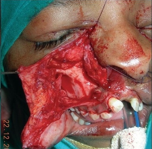
Tessier cleft types 3 and 4 are rare entities even among what are considered other rare craniofacial clefts. Very few cases have been reported worldwide, especially in the bilateral form. In the absence of any well-laid guidelines for management of such rare cases, plastic surgeons operate on such cases due to the inherent complexities in technique. To overcome this problem and provide a ground rule for surgical management of such cases, we propose an easier format with a 'split approach' of the affected areas. In our proposed formatting, we have divided the affected areas of the cleft into three components: 1. Lid component; 2. Lip component; and 3. Nasomalar component. Any person skilled in the plastic surgical art would appreciate that individual management of the aforesaid demarcated areas is easy as compared to the surgery of the entire craniofacial cleft, that too with the contemporary approach. We have evaluated this formatting technique with a 'split approach' in seven cases and found the results more convincing compared to those of classical methods. We invite the surgical fraternity to validate the surgical formatting in their settings and provide us with feedback on the same to consolidate these results.


Conflict of interest statement

Conflict of Interest: None declared.

Figures

Figure 1a
Figure 1a
A 12 year-old girl with bilateral Tessier Cleft type-3
Figure 1b
Figure 1b
3-D CT scan of the same type-3 cleft patient showing severely hypoplastic nasal bone and maintained infra-orbital rim & orbital floor supporting the globe
Figure 2a
Figure 2a
A four year-old boy with Tessier Cleft type-4
Figure 2b
Figure 2b
3-D CT scan of the same type-4 cleft patient with slightly deficient infra-orbital rim & orbital floor; it has a gutter type of appearance (A). There are separate & blind maxillary sinuses which never get a chance to unite with the nasal cavity (B)
Figure 2c
Figure 2c
A case of bilateral Tessier cleft type-4 presented to us at one year of age. The infra-orbital margin & floor of the orbit were deficient (see CT Scan) & the globe was displaced inferiorly to such an extent that the upper eyelid was insufficient to cover the cornea. The cornea was already damaged
Figure 2d
Figure 2d
A case of unilateral Tessier cleft type-4 (wide) at the age of four days. The infra-orbital rim & floor of the orbit is almost completely deficient and is unable to support the globe, which is displaced so inferiorly that the cornea is seen to be already hazy. If no immediate precaution is taken (at least in the form of temporary tarsorrhaphy) to cover the cornea, exposure keratitis & blindness shall surely ensue (Photograph courtesy Dr. Jayachandran, Matha Hospital, Kerala)
Figure 3a
Figure 3a
Simplified marking for Lid & Lip components. For lid component, the incision is marked at the junction of the lower lid & cheek as a back cut and pulled towards the medial canthal area for medial canthopexy. The shaded area of keratinized conjunctiva is to be excised. For Lip component, the incision is marked similar to Veau-III type lip repair with a back cut in the naso-labial crease, maintaining the height of the lip. See Figure 3c for the simplicity of the management of Lid & Lip components after medial canthopexy and Veau-III type lip repair
Figure 3b
Figure 3b
Incision markings described as in Figure 3a. Sometimes, following medial canthopexy, an inferiorly based transposition flap (dotted grey line) from high up in the lateral side of the nose may be required to fill the defect below the lower lid. The shaded area lateral to the philtral column is required to be excised
Figure 3c
Figure 3c
See the simplicity of the surgical steps of Lid & Lip components as after medial canthopexy and Veau-III type lip repair
Figure 3d
Figure 3d
Plane & extent of dissection (subperiosteal and up to the lateral border of the maxilla & zygoma)
Figure 4a
Figure 4a
Markings for the management of the nasomalar component in case of Tessier cleft type-3. See most of the lateral wall of nose (lining) was created by the turn-in flap of cheek tissue which is stitched with a turn-in flap of the lateral nose & alar skin
Figure 4b
Figure 4b
Management of nasomalar component in case of Tessier cleft type-3. See most of the lining of the lateral wall of the nose was created by the turn-in flap of cheek tissue which is stitched with a turn-in flap of the lateral nose & alar skin
Figure 4c
Figure 4c
Closure of the nasomalar component in type-3 cleft after a Z-plasty. Ala was created by rotating & stitching whatever alar tissue was present (worm's eye view)
Figure 4d
Figure 4d
Closure of the nasomalar component in type-3 cleft after a Z-plasty (lateral view)
Figure 5a
Figure 5a
Marking (as described) in B/L Tessier cleft type-4. Shaded area in the upper lip shows zone of excision
Figure 5b
Figure 5b
Dissection of nasomalar component in a case of B/L Tessier cleft type-4. Note the lateral lacrimal duct (going towards & ending in the blind maxillary sinus) and medial lacrimal duct going towards the lateral nasal wall
Figure 5c
Figure 5c
Dissection of B/L Tessier cleft type-4
Figure 5d
Figure 5d
Repair of B/L Tessier cleft type-4 after medial canthopexy & upper lip repair and after application of key stitches
Figure 5e
Figure 5e
After final closure of B/L Tessier cleft type-4. There is usually no requirement of Z-plasty in the nasomalar area and the tissue from the lateral part of the nose naturally falls into the defect created by back-cut of the lip in the nasolabial crease. This tissue is pulled laterally and stitched snugly in the gap of the back-cut
Figure 6
Figure 6
Pre- & postoperative photos of a case of B/L Tessier cleft type-3
Figure 7
Figure 7
Pre- & postoperative photos of a case of B/L Tessier cleft type-4
Figure 8
Figure 8
Adult case of U/L Tessier cleft type-4 with wide gap & depression in the nasomalar area required bone graft from the iliac crest

References

    1. Joos U, Anastassov E. Treatment of craniofacial midline clefts in association with hamartomas: Report of three cases. J Oral Maxillofac Surg. 1998;56:383–92. - PubMed
    1. Ranta R, Rintala R. Oblique lateral oro-ocular facial cleft. J Oral Maxillofac Surg. 1988;17:186–9. - PubMed
    1. Tessier P. Anatomical classification of facial, cranio-facial and latero-facial clefts. J Maxillofac Surg. 1976;4:69–92. - PubMed
    1. Longaker MT, Lipschutz GS, Kawamoto HK. Reconstruction of Tessier No. 4 Clefts Revisited. Plast Reconstr Surg. 1997;99:1501–7. - PubMed
    1. Accessed from: http://www.odv.bo.it/2002-4//04-tessierIV.rtf.