. 2009;4(1):67-78.
doi: 10.1586/17469872.4.1.67.

Nodal as a biomarker for melanoma progression and a new therapeutic target for clinical intervention

Affiliations

Nodal as a biomarker for melanoma progression and a new therapeutic target for clinical intervention

Luigi Strizzi et al. Expert Rev Dermatol. 2009.

Abstract

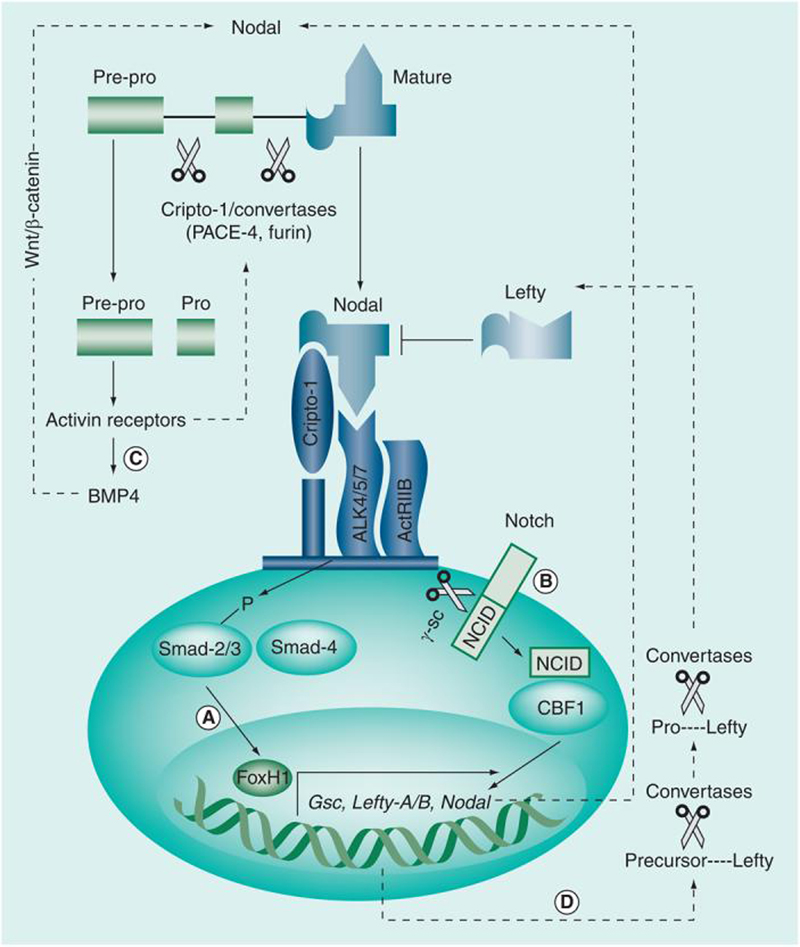
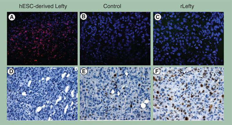
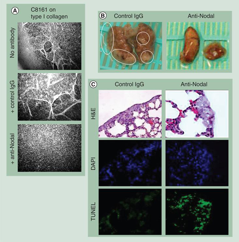
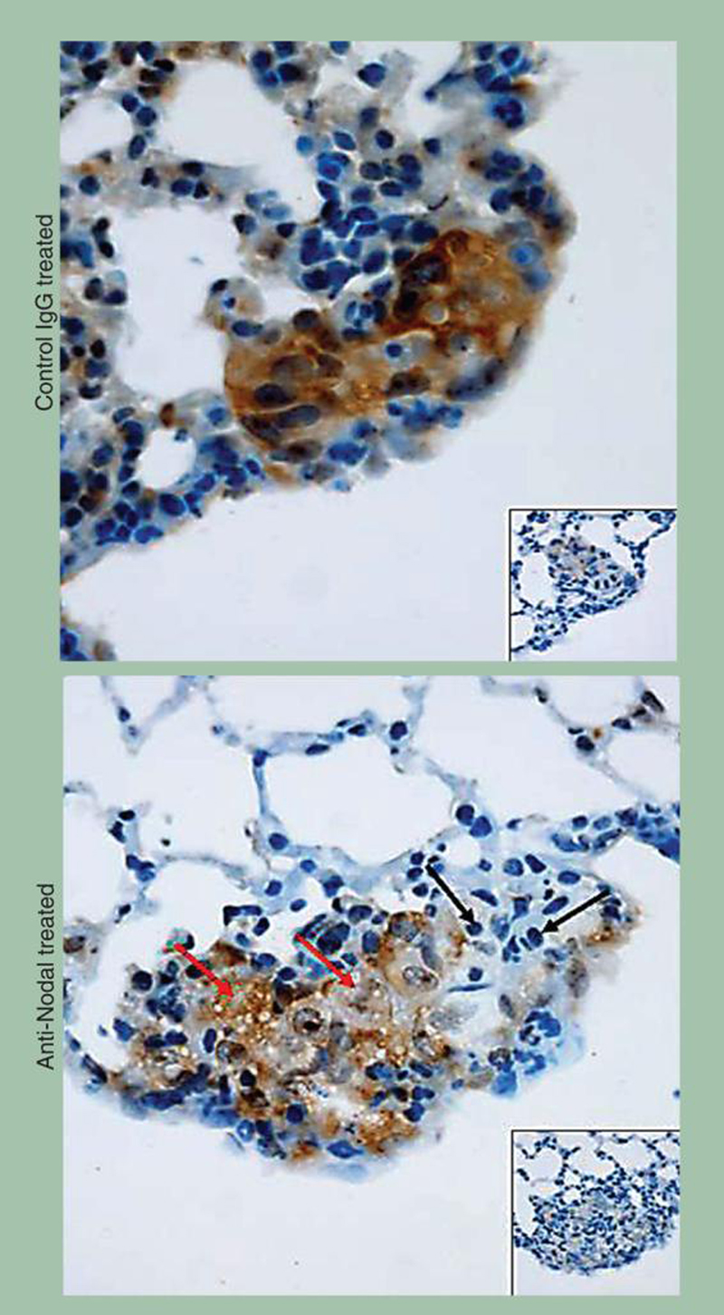
Nodal, an embryonic morphogen belonging to the TGF-β superfamily, is an important regulator of embryonic stem cell fate. We have recently demonstrated that Nodal is expressed significantly in aggressive melanoma. Surprisingly, expression of the Nodal coreceptor, Cripto-1, was detected in only a small fraction of the melanoma tumor cell population, indicating a primary role for Cripto-1-independent signaling of Nodal in melanoma. In this review, we discuss how regulatory factors present in an embryonic environment, such as Lefty, can downregulate Nodal expression and inhibit tumorigenicity and plasticity of melanoma cells. Our translational studies show that antibodies against Nodal are capable of repressing melanoma vasculogenic mimicry and of inducing apoptosis in melanoma tumors in an in vivo lung-colonization assay. Our previous work and ongoing studies suggest that Nodal may represent a novel diagnostic marker and therapeutic target in melanoma.


Figures

Figure 1
Figure 1. Nodal signaling pathways
(A) Classic Nodal signaling involves binding with the coreceptor Cripto-1 and a heteromeric complex composed of Activin type I (ALK 4/5/7) and type II (ActRIIB) serine threonine kinase receptors. Type I and II receptor complexes induce phosphorylation of Smad-2 and -3, which then bind to Smad-4, forming a transcriptional complex that translocates to the nucleus, where it activates the transcription of target genes, such as FoxH1. (B) It is believed that Notch signaling can also induce Nodal expression through binding of the active NICD to CBF1, forming a transcriptional complex that enters the nucleus, inducing transcription of target genes including Nodal. (C) Nodal is synthesized by the cell as pre-pro-Nodal (∼55 kD), which is subject to cleavage by the subtilisin-like proprotein convertases PACE-4 and Furin, which generates the intermediate pro-Nodal (∼37 kD) and eventually active Nodal (∼18 kD). Recent data have suggested that Cripto-1 may play a role during the maturation process of Nodal by recruiting PACE-4/Furin and Nodal to the vicinity of the Nodal receptors on the cell membrane, thereby increasing both efficiency of Nodal processing and availability of the final active form of Nodal to its receptors. It is important to note that immature Nodal is capable of participating in BMP/Wnt/β-catenin signaling pathways, which can also influence Nodal function. Nodal also activates its own transcription via a feed-forward mechanism and the transcription of Lefty. (D) As Lefty exits the cell, it is processed to the active form by specific convertases and acquires the capability of downregulating the function and, ultimately, the expression of Nodal. NCID: Notch intracellular domain.
Figure 2
Figure 2. Depletion of CR-expressing C8161 human melanoma cells and tumorigenicity
(A) C8161 human cutaneous metastatic melanoma cells were depleted of the CR-positive (<5%) subpopulation (red oval), which also expressed higher levels of MDR-1 mRNA, as detected by quantitative real-time PCR (B). The resulting C8161 CR-1-depleted as well as the starting C8161 parental population (control) were then injected subcutaneously into nude mice orthotopically to examine their ability to form tumors. (C) There were no significant differences (Mann–Whitney U test) in tumor volumes between tumors formed by parental C8161 cells and those formed by the CR-depleted C8161 cells, nor were there differences in histological morphology (original magnification 400×). CR: Cripto-1.
Figure 3
Figure 3. Nodal as a biomarker for melanoma progression
(A) Immunohistochemical localization of Nodal (red) in human melanoma. Sections represent RGP melanoma, VGP melanoma and a melanoma metastasis in a lymph node biopsy. Note that RGP melanoma cells express barely detectable Nodal (black arrows) (original magnification 200×). By contrast, strong expression of Nodal is observed in VGP melanoma cells (original magnification 100×) that have invaded subcutaneous connective or adipose tissues (red arrows). Nodal staining is also strong in melanoma cells that have metastasized to the lymph node (original magnification 100×). (B) Detection of Nodal by immunohistochemistry is demonstrated in an example of aggressive human melanoma invading subcutaneous tissue (original magnification 200×). (C) Melanoma cells in advanced VGP melanoma show relatively weak staining for Cripto-1 compared with staining for Nodal from a similar histological area (original magnification 400×). RGP: Radial growth phase; VGP: Vertical growth phase.
Figure 4
Figure 4. Nodal inhibition abrogates aberrant gene expression, invasiveness and tumorigenicity in metastatic melanoma cells.
(A) In vitro invasion assay of C8161 cells cultured in the absence or presence of SB431542 (1 μM or 10 μM), a small molecule inhibitor of Nodal signaling. Invasion was calculated as a percentage of cells able to invade through a defined matrix (collagen IV, laminin and gelatin)-coated membrane during a 24-h period using an in vitro invasion assay. Bars represent the mean normalized invasion indices ± standard deviations. The values indicated by an asterisk are significantly different from the invasion index of control cells (n = 11 wells; p < 0.05, Mann–Whitney U test). Nodal inhibition significantly decreases the invasiveness of C8161 cells. (B) In vivo tumor formation in a mouse injected with C8161 cells treated with either MOControl or MONodal. Values represent the median tumor volume (mm3) ± interquartile range, and the MOControl and MONodal tumor volumes were significantly different at the time points indicated by an asterisk (n = 5; p < 0.05, Mann–Whitney U test). (C) Summary of results from Western blot and reverse transcriptase (RT)-PCR analyses of Nodal, tyrosinase, VE-Cadherin and keratin 8/18 expression in C8161 cells cultured on 3D type I collagen matrices for 6 days in the presence of vehicle (dimethyl sulfoxide), an inhibitor of the Nodal signaling pathway (SB431542, 10 μM) or a Morpholino designed to inhibit Nodal expression (MONodal). Inhibition of Nodal expression resulted in the re-expression of the melanocyte-specific marker tyrosinase concomitant with a reduction in the expression of VE-Cadherin, an endothelial lineage marker, and keratin 8/18, an epithelial lineage marker.
Figure 5
Figure 5. hESC-derived Lefty downregulates Nodal expression and induces apoptosis and decreases proliferation in metastatic melanoma cells in vivo
C8161 human cutaneous metastatic melanoma cells were injected (250,000 cells per animal) orthotopically into a nude mouse model and allowed to form a palpable primary tumor mass for 2 weeks. Subsequently, hESC-derived Lefty (estimated <50 ng), rLefty (30 ng) or carrier minus Lefty (control) was delivered intratumorally over a 2-week period. (A–C) Tumor cell apoptosis was determined by immunohistochemical staining for terminal deoxynucleotidyl transferase biotin–dUTP nick-end labeling (TUNEL), which appears red in the color photomicrographs. (D–F) Immunohistochemistry of the Ki67 proliferation marker appears as a brown staining product. (A) Apoptosis is observed in C8161 melanoma tumor cells injected with Lefty derived from hESCs. (B) No apoptosis is evident in the control tumor receiving a sham injection regime of control or rLefty (C). (D) The proliferation marker Ki67 is not observed in the C8161 melanoma tumors treated with hESC-derived Lefty. (E) Ki67 is abundant in the control tumor receiving a sham injection regime of control or (F) rLefty (original magnification 400×). hESC: Human embryonic stem cell; rLefty: Recombinant Lefty.
Figure 6
Figure 6. Function-blocking anti-Nodal antibody inhibits vasculogenic mimicry in vitro and lung colonization in nude mice by metastatic melanoma cells
(A) C8161 metastatic melanoma cells treated with a function-blocking anti-Nodal antibody were inhibited from engaging in vasculogenic mimicry in vitro compared with no antibody or a nonfunction-blocking IgG control antibody (original magnification 200×). (B & C) C8161 cells were injected retro-orbitally in nude mice and characteristically colonize first to the lung. The mice were then injected intraperitoneally with either a function-blocking anti-Nodal antibody or control IgG over the next 10 days. At the end of this period, the mice were sacrificed and their lungs examined macroscopically, histologically and immunohistochemically. (B) Macroscopic examination of the lungs revealed a significant reduction in the number and size of the tumor cell colonies in the lungs of mice treated with the anti-Nodal antibody compared with the mice treated with the control IgG antibody. (C) Histological examination of H&E-stained lung tissue revealed that the metastatic C8161 cells forming the colonies in the anti-Nodal antibody-treated mice showed evidence of cellular distress characterized by cytoplasmic swelling and vacuolization not observed in the control antibody-treated mice; staining by TUNEL (green stain) showed an increase in the level of apoptosis in the melanoma cells in response to the anti-Nodal antibody compared with the mice treated with the control antibody (original magnification 200×). DAPI staining for cell nuclei was used as a control to locate each cell in the field of view. DAPI: 4′,6-diamidino-2-phenylindole; H&E: Hemotoxylin and eosin; TUNEL: TdT-mediated biotin 16–dUTP nick-end labeling.
Figure 7
Figure 7. Expression of Nodal in lung colonies formed by C8161 human melanoma cells treated with anti-Nodal antibody
Immunohistochemistry shows strong staining for Nodal in the C8161 lung colonies of control IgG-treated mice. Staining for Nodal is less intense in the C8161 lung colonies of mice treated with the anti-Nodal antibody. Of note is the diffuse foam-like appearance of cytoplasmic vacuolization (red arrows) and areas of apoptosis containing evident apoptotic bodies (black arrows) in the anti-Nodal antibody-treated C8161 lung colonies. Insets show tissues stained with irrelevant isotype IgG used as negative control (original magnification 400×).

References

    1. Tsao H, Atkins MB, Sober AJ. Management of cutaneous melanoma. N. Engl. J. Med. 2004;351(10):998–1012. - PubMed
    1. Clark WH, Jr, Elder DE, Guerry D, 4th, et al. A study of tumor progression: the precursor lesions of superficial spreading and nodular melanoma. Hum. Pathol. 1984;15(12):1147–1165. - PubMed
    1. Moan J, Porojnicu AC, Dahlback A. Ultraviolet radiation and malignant melanoma. Adv. Exp. Med. Biol. 2008;624:104–116. - PubMed
    1. Collisson EA, De A, Suzuki H, Gambhir SS, Kolodney MS. Treatment of metastatic melanoma with an orally available inhibitor of the Ras–Raf–MAPK cascade. Cancer Res. 2003;63(18):5669–5673. - PubMed
    1. Haluska F, Pemberton T, Ibrahim N, Kalinsky K. The RTK/RAS/BRAF/PI3K pathways in melanoma: biology, small molecule inhibitors, and potential applications. Semin. Oncol. 2007;34(6):546–554. - PubMed

Website

    1. The Cancer Genome Anatomy Project: SAGE Anatomic Viewer (SAV) http://cgap.nci.nih.gov/SAGE/AnatomicViewer.

LinkOut - more resources