. 2010 Feb;222(2):387-400.
doi: 10.1002/jcp.21963.

Integrin-linked kinase regulates migration and proliferation of human intestinal cells under a fibronectin-dependent mechanism

Affiliations
Free PMC article

Integrin-linked kinase regulates migration and proliferation of human intestinal cells under a fibronectin-dependent mechanism

David Gagné et al. J Cell Physiol. 2010 Feb.
Free PMC article

Abstract

Integrin-linked kinase (ILK) plays a role in integrin signaling-mediated extracellular matrix (ECM)-cell interactions and also acts as a scaffold protein in functional focal adhesion points. In the present study, we investigated the expression and roles of ILK in human intestinal epithelial cells (IECs) in vivo and in vitro. Herein, we report that ILK and its scaffold-function interacting partners, PINCH-1, alpha-parvin, and beta-parvin, are expressed according to a decreasing gradient from the bottom of the crypt (proliferative/undifferentiated) compartment to the tip of the villus (non-proliferative/differentiated) compartment, closely following the expression pattern of the ECM/basement membrane component fibronectin. The siRNA knockdown of ILK in human IECs caused a loss of PINCH-1, alpha-parvin, and beta-parvin expression, along with a significant decrease in cell proliferation via a loss of cyclin D1 and an increase in p27 and hypophosphorylated pRb expression levels. ILK knockdown severely affected cell spreading, migration, and restitution abilities, which were shown to be directly related to a decrease in fibronectin deposition. All ILK knockdown-induced defects were rescued with exogenously deposited fibronectin. Altogether, our results indicate that ILK performs crucial roles in the control of human intestinal cell and crypt-villus axis homeostasis-especially with regard to basement membrane fibronectin deposition-as well as cell proliferation, spreading, and migration.


Figures

Fig. 1
Fig. 1
Expression of ILK, PINCH1, α-parvin, and fibronectin in the human small intestine. Immunolocalization on human intestinal cryosections of (A) ILK, (B) PINCH-1, (C) α-parvin, and (D) fibronectin with specific antibodies showed strong staining in the basal membrane of the epithelial and mesenchymal compartments along the crypt. Parts A–D are a composite of adjacent areas. Immunolocalization of (E) ILK and (E′) the laminin chains alone (small arrows) or (E′′) together confirmed the localization of ILK at the epithelial basal membrane (large arrows). F: Semi-quantitative RT-PCR of ILK, PINCH-1, α-parvin, and β-parvin on intestinal epithelial and mesenchymal fractions showed epithelial and mesenchymal expression for each protein. Vimentin and E-cadherin were used to control for fraction purity, and RPLPO was used as a normalizing gene. NTC, no template control; c, crypts; v, upper half of the villus. A–D: Scale bar in (A): 50 µm; in (E–E″), scale bar in (E): 20 µm.
Fig. 2
Fig. 2
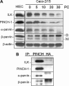
Regulation of P1IP complex members during proliferation in differentiating intestinal epithelial cells. A: Representative Western blot analyses of whole cell lysates of HIEC and Caco-2/15 cells at different stages of post-confluence (0–30 days) using specific antibodies directed against members of the P1IP complex. ILK, PINCH-1, α-parvin, and β-parvin were expressed in our in vitro models. PINCH-1, ILK, α-parvin, and β-parvin(s) (lower band) expression decreased during Caco-2/15 differentiation. β-parvin(l) (upper band) increased during the differentiation process. β-actin was used as a loading control. Representative Western analysis of experiments carried out at least three times independently. B: Co-IP of the P1IP members using PINCH antibody and HA-probe antibody as control on HIEC cells. Immunoblot analysis shows co-immunoprecipitation of ILK, α-parvin, and two isoforms of β-parvin with PINCH-1. Representative Western analysis of experiments carried out three times independently. PC, post-confluence.
Fig. 3
Fig. 3
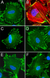
Localization of P1IP members at focal adhesion sites in HIEC cells. Cells were plated on glass coverslips, fixed, and stained with different specific antibodies against (A) vinculin (green) or (B) vinculin (green) co-localized with F-actin (red), (C) V5-ILK, (D) PINCH-1, (E) α-parvin, and (F) β-parvin. Staining patterns were typical of focal adhesion points. A–F: Scale bar in (A): 10 µm. Representative images of experiments carried out at least three times independently.
Fig. 4
Fig. 4
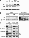
siRNA inhibition of ILK expression in IEC cells. A: HIEC cells were transfected with 10, 25, or 100 µM of siRNA directed against ILK (siILK), 100 µM of a non-silencing negative control siRNA (siCNS), or without siRNA (WT) and monitored for ILK expression. Western analysis for FAK and β-actin protein levels was used as controls for siRNA specificity and loading, respectively. B: HIEC and Caco-2/15 cells transfected with 40 µM of siILK or the non-silencing negative control siCNS were analyzed by Western blot for ILK expression as a function of time (HIEC, 4–10 days; Caco-2/15, 5 and 8 days). β-actin was used as a loading control. C: HIEC and Caco-2/15 cells were transfected with 40 µM siILK, harvested after 72 h (Caco-2/15 cells were at 0 days PC), and analyzed by Western blot using specific antibodies against ILK, PINCH-1, α-parvin, and β-parvin. β-actin was used as a loading control. siILK caused a decrease in expression of all P1IP components in both cell types. Representative Western analysis of experiments carried out at least three times independently. WT, wild type.
Fig. 5
Fig. 5
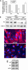
Knockdown of ILK expression inhibits IEC cell fibronectin deposition. HIEC cells were transfected with 40 µM of siCNS or siILK. A: Real-time PCR was carried out on HIEC cells to measure levels of ILK, fibronectin (FN), and laminin β1-chain (LNβ) mRNA levels in siCNS-transfected cells (gray columns) versus silk-transfected cells (white columns). siILK cells showed a significant decrease in ILK and fibronectin mRNA levels. Results are expressed as mean ± standard error of the mean (SEM) and are from three independent experiments. ***P < 0.001 in a one-sample t-test with the hypothetical mean of 100 for the normalized values of siCNS. B: Protein levels of fibronectin and the laminin β1γ1-chains were analyzed in the medium, in the insoluble matrix, and in whole cell lysates of HIEC siCNS and siILK cells, and a decrease of fibronectin levels was observed in siILK cell insoluble matrix and whole cell lysates. Representative Western images from five independent experiments. Indirect immunofluorescence of insoluble fibronectin on HIEC (C) siCNS and (D) siILK cells. C,D: Scale bar in (C): 10 µm. E: Representative Western blot analysis of the α5 subunit of the α5β1 integrin and αV subunit of the αV-containing integrins from siCNS- and siILK-treated HIEC showing no significant variation in three separate experiments. [Color figure can be viewed in the online issue, which is available at www.interscience.wiley.com.]
Fig. 6
Fig. 6
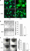
Knockdown of ILK expression inhibits HIEC cell spreading and migration and Caco-2/15 monolayer restitution. HIEC or Caco-2/15 cells were transfected with 40 µM of siCNS or siILK. A: Transfected HIEC cells were plated on plastic and observed at 4 and 18 h, fixed, and stained for vinculin. Scale bar: 20 µm. B: siRNA-treated HIEC were grown on plastic until confluence and wounded with a razor blade. Cells which had migrated into the wounded area after 48 h were counted and expressed as a percentage of control cells. Results are expressed as mean ± standard error of the mean (SEM) and are from three independent experiments. **P < 0.01 in a one-sample t-test with the hypothetical mean of 100 for the normalized values of siCNS. C: siRNA-treated Caco-2/15 cells were grown until confluence and the monolayer was wounded by aspiration. Original wound and remaining wound areas where cells had not closed the original wound after 48 h were measured and the results are expressed as a percentage of the restitution of the original wounded area. Results are expressed as mean ± standard error of the mean (SEM) and are from three independent experiments. *P < 0.05 in a paired t-test. Scale bar: 100 µm. [Color figure can be viewed in the online issue, which is available at www.interscience.wiley.com.]
Fig. 7
Fig. 7
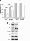
Knockdown of ILK expression inhibits HIEC cell proliferation. HIEC and Caco-2/15 cells were transfected with 40 µM of siCNS or siILK. A: Proliferation indexes were determined by BrdU incorporation on transfected cells that were seeded on plastic or on exogenous fibronectin, and stained using an anti-BrdU antibody. BrdU-positive cells were counted and expressed as a percentage of total cells determined by DAPI staining. Results are expressed as mean ± standard error of the mean (SEM) and are from three independent experiments. *P < 0.05 in a paired t-test. B: HIEC-transfected cells were analyzed by Western blot for protein expression levels of ILK, pRB (upper band, hyperphosphorylated form; lower band, hypophosphorylated form), cyclin D, p21, and p27. β-actin was used as a loading control. Representative Western analysis is from at least three independent experiments. n.s., not significant.
Fig. 8
Fig. 8
Exogenous fibronectin rescued spreading, migration, and restitution of siILK cells. HIEC or Caco-2/15 cells were transfected with 40 µM of siCNS or siILK. A: HIEC cell aggregates (50–100 cells) were plated on plastic or exogenous fibronectin and analyzed after 24 h. The area covered by the spread/migrated cells was subsequently measured and the relative measure of spreading and migration was established by calculating the ratios of the area covered by cells from the aggregates over the total number of cells in the spread aggregate compared to controls × 100 (expressed as “% of control”). Results are expressed as mean ± standard error of the mean (SEM) and are from at least three independent experiments. ***P < 0.001 in a one-sample t-test with the hypothetical mean of 100 for the normalized values of siCNS. B: siRNA-treated Caco-2/15 cells were grown until confluence and the monolayers were wounded by aspiration. Exogenous fibronectin was then added or not to the culture medium. Areas where cells had not covered the original wound after 48 h were measured and expressed as a percentage of the original wounded area. Results are expressed as mean ± standard error of the mean (SEM) and are from at least three independent experiments. *P < 0.05; ***P < 0.001 in a paired t-test. C: siILK- and siCNS-treated HIEC cells were plated and grown on plastic or fibronectin for 48 h. The cells were then harvested and analyzed by Western blot. Fibronectin did not rescue the decrease of the PIP components caused by siILK treatment. β-actin was used as a loading control. n.s., not significant.
Fig. 9
Fig. 9
Knockdown of ILK expression does not influence the differentiation process. Caco-2/15 cells were transfected with 40 µM of siCNS or siILK. Real-time PCR was carried out to measure DPPIV, villin, and sucrase–isomaltase (SI) mRNA levels in siCNS cells (gray columns) versus siILK cells (white columns). Abolition of ILK in Caco-2/15 cells did not significantly change DPPIV, villin, and SI transcript levels at 6 days PC. Results are expressed as mean ± SEM and are from three independent experiments. Statistical analysis was one-sample t-test with the hypothetical mean of 100 for the normalized values of siCNS. n.s., not significant; PC, post-confluence.

References

    1. Alonso CR, Pesce CG, Kornblihtt AR. The CCAAT-binding proteins CP1 and NF-I cooperate with ATF-2 in the transcription of the fibronectin gene. J Biol Chem. 1996;271:22271–22279. - PubMed
    1. Assi K, Mills J, Owen D, Ong C, St Arnaud R, Dedhar S, Salh B. Integrin-linked kinase regulates cell proliferation and tumour growth in murine colitis-associated carcinogenesis. Gut. 2008;57:931–940. - PubMed
    1. Baneyx G, Baugh L, Vogel V. Fibronectin extension and unfolding within cell matrix fibrils controlled by cytoskeletal tension. Proc Natl Acad Sci USA. 2002;99:5139–5143. - PMC - PubMed
    1. Basson MD. In vitro evidence for matrix regulation of intestinal epithelial biology during mucosal healing. Life Sci. 2001;69:3005–3018. - PubMed
    1. Beaulieu JF. Differential expression of the VLA family of integrins along the crypt-villus axis in the human small intestine. J Cell Sci. 1992;102:427–436. - PubMed

Publication types

MeSH terms