Review
. 2010 Jan;31(1):9-18.
doi: 10.1093/carcin/bgp268. Epub 2009 Nov 3.

Telomeres and telomerase in cancer

Affiliations
Review

Telomeres and telomerase in cancer

Steven E Artandi et al. Carcinogenesis. 2010 Jan.

Abstract

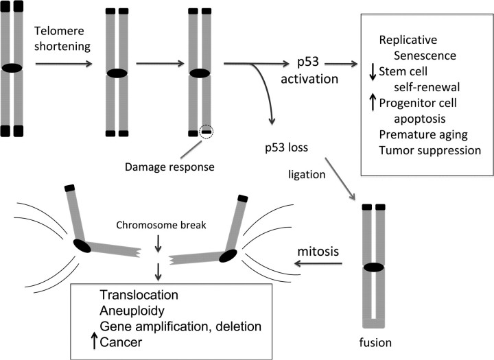
Myriad genetic and epigenetic alterations are required to drive normal cells toward malignant transformation. These somatic events commandeer many signaling pathways that cooperate to endow aspiring cancer cells with a full range of biological capabilities needed to grow, disseminate and ultimately kill its host. Cancer genomes are highly rearranged and are characterized by complex translocations and regional copy number alterations that target loci harboring cancer-relevant genes. Efforts to uncover the underlying mechanisms driving genome instability in cancer have revealed a prominent role for telomeres. Telomeres are nucleoprotein structures that protect the ends of eukaryotic chromosomes and are particularly vulnerable due to progressive shortening during each round of DNA replication and, thus, a lifetime of tissue renewal places the organism at risk for increasing chromosomal instability. Indeed, telomere erosion has been documented in aging tissues and hyperproliferative disease states-conditions strongly associated with increased cancer risk. Telomere dysfunction can produce the opposing pathophysiological states of degenerative aging or cancer with the specific outcome dictated by the integrity of DNA damage checkpoint responses. In most advanced cancers, telomerase is reactivated and serves to maintain telomere length and emerging data have also documented the capacity of telomerase to directly regulate cancer-promoting pathways. This review covers the role of telomeres and telomerase in the biology of normal tissue stem/progenitor cells and in the development of cancer.


Figures

Fig. 1.
Fig. 1.
Telomeres protect chromosome ends. Telomeres are TTAGGG double-stranded DNA repeats culminating in a single-stranded overhang. The shelterin protein complex protects telomeres, facilitates replication and controls access of telomerase. TRF1 and TRF2 bind the double-stranded telomere. POT1 and TPP1 are OB-fold containing proteins associated with the single stranded overhang. RAP1 binds TRF2, and TIN2 is a central component of the complex interacting with TRF1, TRF2 and TPP1. Telomeres can exist in a looped conformation, termed the t-loop, in which the single-stranded overhang folds back on the double-stranded telomere to sequester the end. The 3′ hydroxyl group represents the substrate for telomere addition by telomerase.
Fig. 2.
Fig. 2.
Telomere shortening activates p53 and drives formation of epithelial cancers through gene amplification and deletion. Telomeres shorten progressively with cell division due to the end-replication problem in settings of insufficient telomerase, including in human fibroblasts, aging tissues, early cancers and diseases of high cellular turnover. Critical telomere shortening compromises the telomere cap and results in a DNA damage response that activates the p53 tumor suppressor protein. This activation of p53 induces replicative senescence in cultured human fibroblasts, impairs stem cell self-renewal, induces apoptosis in tissue progenitor cells, causes premature aging and strongly suppresses tumor formation. If p53 is mutated or deleted, these responses to telomere dysfunction are mitigated and chromosomal fusions are tolerated. The generation of fused chromosomes results in dicentric chromosomes (chromosomes with two centromeres) and when these attach to opposite spindle poles, chromosome breakage occurs. These broken ends serve as potent catalysts for translocations, focal amplifications and focal deletions. Such CNAs drive development of carcinomas and explain the widespread gene copy number changes seen in human cancers.
Fig. 3.
Fig. 3.
Telomerase is a large multisubunit RNP. Telomerase is a reverse transcriptase that adds telomere repeats to chromosome ends. The minimal catalytic core is composed of TERT, the telomerase reverse transcriptase, and TERC, the telomerase RNA, which acts as the template for telomere addition. The 5′ end of TERC contains the template region (black box) and 3′ end of TERC contains two sequences that act as binding sites for additional telomerase protein factors. The H/ACA box represents the binding site for dyskerin, a protein critical for telomerase assembly and for stability of TERC. Dyskerin has three small associated proteins—NHP2, NOP10 and GAR1—shown as blue spheres. TCAB1 is a WD40 repeat protein that recognizes the CAB box in TERC. TCAB1 interacts with dyskerin and is crucial for facilitating telomerase trafficking to Cajal bodies and for telomere maintenance.

References

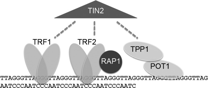
    1. Palm W, et al. How shelterin protects mammalian telomeres. Annu. Rev. Genet. 2008;42:301–334. - PubMed
    1. Griffith JD, et al. Mammalian telomeres end in a large duplex loop. Cell. 1999;97:503–514. - PubMed
    1. van Steensel B, et al. TRF2 protects human telomeres from end-to-end fusions. Cell. 1998;92:401–413. - PubMed
    1. Celli GB, et al. DNA processing is not required for ATM-mediated telomere damage response after TRF2 deletion. Nat. Cell Biol. 2005;7:712–718. - PubMed
    1. van Steensel B, et al. Control of telomere length by the human telomeric protein TRF1. Nature. 1997;385:740–743. - PubMed