. 2009 Dec 1;122(Pt 23):4303-10.
doi: 10.1242/jcs.057646. Epub 2009 Nov 3.

Crucial role of phosphatidylinositol 4-kinase IIIalpha in development of zebrafish pectoral fin is linked to phosphoinositide 3-kinase and FGF signaling

Affiliations

Crucial role of phosphatidylinositol 4-kinase IIIalpha in development of zebrafish pectoral fin is linked to phosphoinositide 3-kinase and FGF signaling

Hui Ma et al. J Cell Sci. .

Abstract

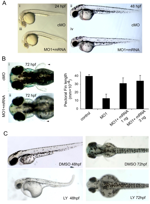
Phosphatidylinositol 4-kinases (PI4Ks) catalyze the first committed step in the synthesis of phosphoinositides, important lipid regulators of signaling and trafficking pathways. Here we cloned Pik4a, one of the zebrafish PI4K enzymes, and studied its role(s) in vertebrate development using morpholino oligonucleotide-based gene silencing in zebrafish. Downregulation of Pik4a led to multiple developmental abnormalities, affecting the brain, heart, trunk and most prominently causing loss of pectoral fins. Strikingly similar defects were caused by treatment of the developing embryos with the phosphoinositide 3-kinase (PI3K) inhibitor, LY294002. To investigate the cause of the pectoral fin developmental defect, we focused on fibroblast growth factor (FGF) signaling pathways because vertebrate limb development requires the concerted action of a series of FGF ligands. Using in situ hybridization, the pectoral fin defect was traced to disruption of the early FGF signaling loops that are crucial for the establishment of the sharp signaling center formed by the apical ectodermal ridge and the underlying mesenchyme. This, in turn caused a prominent loss of the induction of one of the mitogen-activated protein kinase (MAPK) phosphatases, Mkp3, an essential intermediate in vertebrate limb development. These changes were associated with impaired proliferation in the developing fin bud due to a loss of balance between the MAPK and PI3K branch of FGF-initiated signals. Our results identify Pik4a as an upstream partner of PI3Ks in the signaling cascade orchestrated by FGF receptors with a prominent role in forelimb development.


Figures

Fig. 1.
Fig. 1.
Expression and downregulation of zebrafish Pi4ka. (A) Pi4ka cloned from zebrafish shows high homology to other mammalian Pi4ka enzymes sharing the same domain organization and highest conservation within the C-terminal catalytic domain. (B) Expression pattern of pi4ka mRNA during zebrafish embryogenesis. Expression is ubiquitous in early embryos (1.5-24 hpf) but is primarily restricted to the brain, branchial arches (ba), and fin buds (fb, indicated by arrowheads) at later stages (36 and 48 hpf). (C) Downregulation of zebrafish Pi4ka by morpholino injection targeting the splicing of exon 50 that encodes a crucial region within the catalytic domain (MO1 and MO2). Left panels: RT-PCR analysis showed that antisense morpholinos could eliminate the transcript containing exon 50. Both MOs caused exon skipping in a dose-dependent manner, whereas control injections (FITC) were without effect. Right panel: in vitro translation assay showing the ability of MO3 and MO4 targeting the translation initiation site to reduce in vitro translation of Pi4ka (cMO, control morpholino). (D) Lateral view of MO1 (9 ng)-injected morphants show a complex phenotype affecting several structures. Note the shorter trunk with curved tail, smaller head and eye, reduced and disorganized pigmentation, and pericardial edema. Dorsal view of the embryos at 72 hpf, demonstrate the larger yolk sac, smaller head and eyes, and the loss of pectoral fins (pf, arrowheads) in morphants.
Fig. 2.
Fig. 2.
pi4ka mRNA co-injection can reverse, and PI3K inhibitors can mimic the MO1-induced phenotype. Fertilized zebrafish eggs were injected at the one-cell stage with either a control morpholino (cMO) or MO1 together with in vitro transcribed and purified pi4ka mRNA. Embryos were allowed to develop for 1-3 days (24-72 hpf). MO1 interferes with splicing of the endogenous mRNA causing exon skipping but does not affect the translation of the injected full length mRNA. (A) Co-injection of 1 ng mRNA can largely alleviate the brain defect at 24 hpf and 48 hpf. (B) The pectoral fin develops to almost normal length, but it poorly separates from the yolk sac and does not fully develop in the rescued embryos (left panels; arrowhead). Measurements of the length of the pectoral fin at the 72 hpf stage (right panel) show partial rescue by the injected mRNA (values are means ± s.e.m. of 70-110 observations). (C) The PI3K inhibitor, LY294002 (10 μM) mimics the effects of pi4ka downregulation on zebrafish development. LY-treated embryos have a smaller brain and eyes, a hooked tail and pericardial edema at 48 hpf. At 72 hpf, LY-treated embryos have larger yolk sacs and also reduced pectoral fins. These changes are very similar to those observed in pi4ka downregulated embryos (see Fig. 1D).
Fig. 3.
Fig. 3.
Effect of pi4ka downregulation and LY294002 treatment expression of several genes on the early FGF signaling pathway. Fertilized zebrafish eggs were injected at the one-cell stage either with a control morpholino (cMO) or with MO1 or treated with 10 μM LY294002 and analyzed by whole-mount in situ hybridization for the presence of the indicated mRNA at different stages of development. (A) At 24 hpf embryos mkp3 expression is reduced in the fin-bud mesenchyme in the morphants as well as in embryos treated with 10 μM LY294002. This difference becomes very prominent by the 36 hpf stage and also affects the branchial arches. (B) Expression of tbx5 is only slightly reduced in the fin-bud mesenchyme of morphant embryos both at 24 and 32 hpf. The expression of fgf24 is comparable to controls in the fin-bud mesenchyme of pi4ka morphant embryos at 24 hpf. However, at 32 hpf, fgf24-positive cells move to the AER in control embryos but are still dispersed in the mesenchyme and fail to migrate to the AER in pi4ka morphant embryos. As a result, expression of fgf10, which is controlled by fgf24 in the AER, is greatly reduced in the fin-bud mesenchyme of Pi4ka morphant embryos. Note that some signals in the control are saturated because identical exposures were chosen to still see the weak signal in the morphant embryos.
Fig. 4.
Fig. 4.
Fluorescent in situ hybridization (FISH) and confocal analysis of fgf24 and mkp3 expression and localization. (A) Time sequence of the position of the fgf24-positive cells showing their migration to the AER that is very clear by 36 hpf. At the same time, the number of mkp3-positive cells and their signal progressively increases but remains beneath the AER. The DAPI staining is not shown for better clarity but the contours of the ectoderm based on the DAPI staining are indicated by a dashed line (see panel B). (B) Representative images showing the position of the developing fin bud in a lateral view of a control fish embryo at the 36 hpf stage analyzed by FISH for mkp3 (orange) and counterstained with DAPI (green). The boxed region of each image is enlarged in the panel to the right. (C) Left: schematic illustration of the interplay between the AER and the underlying mesenchyme showing the position of fgf24- and mkp3-positive cells at the 30 hpf stage. Right: at the 36 hpf stage the fgf24-expressing cells do not approach the apical layer of the budding fin and the AER is not developing. As a consequence the mkp3 expression remains weak and the cells are scattered in the pi4ka MO1-injected and LY294002-treated embryos. Note that these pictures do not reflect the true relative signals in the control and morphant embryos, because the images were adjusted to make the position of the positive cells clear. LPM, lateral plate mesenchyme.
Fig. 5.
Fig. 5.
Loss of Pi4ka function leads to widespread cell death and reduced cell proliferation. (A) Cell death detected by TUNEL analysis showed few positive cells in control embryos (cMO) at 24 hpf (i, iii) and 36 hpf stage (v). Embryos injected with pi4ka MO1 had large numbers of apoptotic cells throughout the embryo at both 24 and 36 hpf stages especially in the brain, eyes and the trunk. Notably, there was significant cell death in the lateral plate mesoderm region (iv, vi) the early precursor of the pectoral fin bud. Acridine orange (AO) staining (vii-x) showed similar pattern of apoptosis at 24 hpf in pi4ka MO1-injected (vii versus viii) or LY294002-treated (ix versus x) embryos. (B) Proliferating cells were detected by immunohistochemistry using anti-H3P antibody. Proliferation was significantly lower in the brain of pi4ka MO1-injected embryos (MO1) than in that of controls (cMO). Proliferation was also greatly reduced at the somites and lateral plate mesenchyme at 24 hpf and almost completely disappeared in the pectoral fin buds region of pi4ka morphants at 48 hpf. (C) H3P-positive cells in the mesenchyme of fin buds at 48 hpf were quantified.Values are means ± s.d. of at least 20 measurements on independent embryos.
Fig. 6.
Fig. 6.
Schematics of the proposed participation of Pi4ka and PI3Ks in the FGF signaling cascade in the developing fin bud mesenchyme. Pi4ka contributes to the generation of the PtdIns(4,5)P2 pool that is converted by the PI3Ks to PtdIns(3,4,5)P3 in the plasma membrane. Production of this latter lipid activates the Akt signaling cascade that regulates a host of downstream genes important for proliferation or protection of the cells from apoptosis. One of the most important genes regulated by this pathway is the MAPK phosphatase, MKP3 that is a key intermediate in driving pectoral fin development (Kawakami et al., 2003). This phosphatase keeps a right balance between MAPK and the Akt pathways. The right panel shows the FGF signaling cascade in which the Fgf24-positive cell migration appears to be the first step showing impairment after Pi4ka depletion.

References

    1. Ashworth, R., Devogelaere, B., Fabes, J., Tunwell, R. E., Koh, K. R., De Smedt, H. and Patel, S. (2007). Molecular and functional characterization of inositol trisphosphate receptors during early zebrafish development. J. Biol. Chem. 282, 13984-13993. - PubMed
    1. Audhya, A. and Emr, S. D. (2002). Stt4 PI 4-kinase localizes to the plasma membrane and functions in the Pkc1-mediated MAP kinase cascade. Dev. Cell 2, 593-605. - PubMed
    1. Balla, A. and Balla, T. (2006). Phosphatidylinositol 4-kinases; old enzymes with emerging functions. Trends Cell Biol. 16, 351-361. - PubMed
    1. Balla, A., Kim, Y. J., Varnai, P., Szentpetery, Z., Knight, Z., Shokat, K. M. and Balla, T. (2007). Maintenance of hormone-sensitive phosphoinositide pools in the plasma membrane requires phosphatidylinositol 4-kinase III{alpha}. Mol. Biol. Cell 19, 711-721. - PMC - PubMed
    1. Balla, A., Tuymetova, G., Toth, B., Szentpetery, Z., Zhao, X., Knight, Z. A., Shokat, K., Steinbach, P. J. and Balla, T. (2008). Design of drug-resistant alleles of type-III phosphatidylinositol 4-kinases using mutagenesis and molecular modeling. Biochemistry 47, 1599-1607. - PubMed

Publication types

MeSH terms

Substances