. 2009 Dec 1;69(23):9038-46.
doi: 10.1158/0008-5472.CAN-09-1499. Epub 2009 Nov 3.

Epigenetic repression of microRNA-129-2 leads to overexpression of SOX4 oncogene in endometrial cancer

Affiliations

Epigenetic repression of microRNA-129-2 leads to overexpression of SOX4 oncogene in endometrial cancer

Yi-Wen Huang et al. Cancer Res. .

Abstract

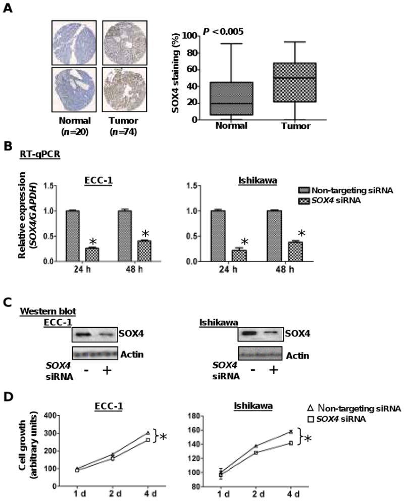
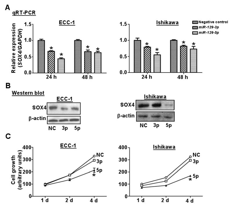
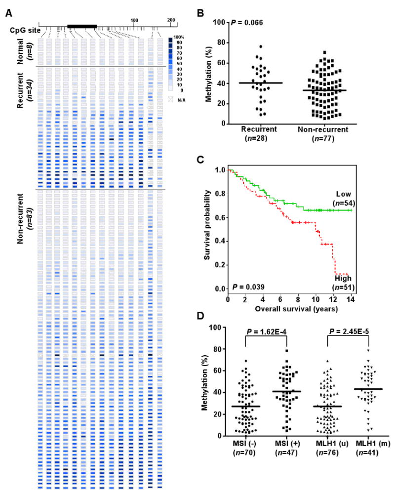
Genetic amplification, mutation, and translocation are known to play a causal role in the upregulation of an oncogene in cancer cells. Here, we report an emerging role of microRNA, the epigenetic deregulation of which may also lead to this oncogenic activation. SOX4, an oncogene belonging to the SRY-related high mobility group box family, was found to be overexpressed (P < 0.005) in endometrial tumors (n = 74) compared with uninvolved controls (n = 20). This gene is computationally predicted to be the target of a microRNA, miR-129-2. When compared with the matched endometria, the expression of miR-129-2 was lost in 27 of 31 primary endometrial tumors that also showed a concomitant gain of SOX4 expression (P < 0.001). This inverse relationship is associated with hypermethylation of the miR-129-2 CpG island, which was observed in endometrial cancer cell lines (n = 6) and 68% of 117 endometrioid endometrial tumors analyzed. Reactivation of miR-129-2 in cancer cells by pharmacologic induction of histone acetylation and DNA demethylation resulted in decreased SOX4 expression. In addition, restoration of miR-129-2 by cell transfection led to decreased SOX4 expression and reduced proliferation of cancer cells. Further analysis found a significant correlation of hypermethylated miR-129-2 with microsatellite instability and MLH1 methylation status (P < 0.001) and poor overall survival (P < 0.039) in patients. Therefore, these results imply that the aberrant expression of SOX4 is, in part, caused by epigenetic repression of miR-129-2 in endometrial cancer. Unlike the notion that promoter hypomethylation may upregulate an oncogene, we present a new paradigm in which hypermethylation-mediated silencing of a microRNA derepresses its oncogenic target in cancer cells.


Figures

Figure 1
Figure 1
SOX4 is overexpressed in endometrial tumors. A, representative photographs of endometrial tissue microarrays (1.5mm core diameter) which underwent immunohisochemical staining for SOX4 and were scored for nuclear staining by the TissueMine software. Boxplots of SOX4 nuclear staining for normal tissue (n=20) and endometrial tumors (n=74) P < 0.005. B & C, relative expression levels of SOX4 mRNA and protein in ECC-1 and Ishikawa cells after transient transfection with SOX4 siRNA or a pool of non-targeting siRNA oligonucleotides for 24 and/or 48 h. GADPH and β-actin served as internal controls for RT-qPCR and western blotting respectively. Error bars represent SD among triplicates; *, P<0.05. D, cellular proliferation was measured by MTS assay in endometrial cancer cells transfected with SOX4 siRNA or non-targeting siRNA. Transfectants (3,000/well) were placed in 96-well plates and proliferation was measured every 24 h. Each point represents the mean value of at least triplicates. *, P < 0.05.
Figure 2
Figure 2
miR-129-2 directly targets SOX4. A, bioinformatic analysis of miR-129-3 and -5p (the mature forms of miR-129-2) predicted binding sites in the SOX4 3′-UTR (bars under line). The predicted pairing of mRNA target region (top) and miRNA (bottom) is as indicted, where a line (“|”) indicates hydrogen bonding. B, predicted secondary structure of miR-129-2. The stem loop structure of miR-129-2 is a precursor to two mature miRNAs: miR-129-3p, and miR-129-5p (bold text). C, miR-129-3p and -5p suppressed the expression of a luciferase vector with the SOX4 3′-UTR. A luciferase expression vector with the 3′-UTR of SOX4 and UBE2F or an empty vector was transfected into ECC-1 and Ishikawa cells. At the same time, anti-miR-129-3p or -5p (anti-3p or anti-5p) and/or miR-129-3p or 5p (3p or 5p) were introduced. Twenty-four hours after the transfection, the cells were harvested and assayed for luciferase activity. Renilla luciferase was used for normalization to empty vector for the transfection efficiency. Error bars, SD; *, P < 0.05 compared with empty vector. D, dot plots showing an inverse relationship between SOX4 (left) and miR-129-3p (right) mRNA expression in 31 pairs of endometrial tumors and adjacent normal tissues. Horizontal bars represent mean expression levels. Significant differences were determined using Student's t tests.
Figure 3
Figure 3
Reactivation of miR-129-2 in cancer cells by pharmacological induction of hyperacetylation and DNA demethylation lead to reduced SOX4 expression at both the mRNA and protein levels. A, genomic map of miR-129-2 CpG island and amplicon. Bar under line, CpG site; ↓, AciI cutting sites. B, COBRA analysis in endometrial cancer cell lines. u, unmethylated band; m, methylated bands; SssI, 100% methylated control; Blood, a mix of 4 normal peripheral blood samples as negative control; +, AciI restriction enzyme added; -, without AciI. C, relative expression levels of miR-129-3p in endometrial cancer cell lines treated with DAC and/or TSA in relation to untreated controls was determined by RT-qPCR analysis. RNU48 was used as internal control gene. Error bar, SD; *, P < 0.05 compared with untreated control of the same cell type. D, relative expression levels of SOX4 mRNA (right) and protein (right) indicating a down-regulation of SOX4 in endometrial cancer cells after treatment with DAC and/or TSA. GAPDH or β-actin was used as internal or loading control, respectively.
Figure 4
Figure 4
Functional analysis of miR-129-2 in endometrial cancer cell lines. A & B, relative levels of SOX4 mRNA (A) or protein (B) expression in ECC-1 and Ishikawa cells after transient transfection with miRNAs or negative control (NC) RNA oligonucleotides for 24 or 48 h. GADPH and β-actin served as an internal control of mRNA or protein, respectively. Error bars, SD from triplicates; *, P < 0.05 compared with NC at the same time point. C, cellular proliferation by MTS assay in the endometrial cancer cell lines ECC-1 and Ishikawa transfected with miR-129-3p (3p), miR-129-5p (5p), or negative control (NC). Proliferation measured as described in Fig. 1C.
Figure 5
Figure 5
Methylation of miR-129-2 CpG island and clinic-pathological covariates analyses in primary endometrioid endometrial tumors. A, methylation profiles of 8 normal endometrial tissues, 34 recurrent, and 83 non-recurrent primary tumors created following MassARRAY analysis. Each row represents a sample and each column represents a CpG unit. Color-coding depicts the degree of methyltion with dark blue being 100% and white being 0%; N/A, not analyzable. B, dot plots point that miR-129-2 hypermethylation is moderately correlated with recurrent diseases. The mean of normal specimens in Fig. 5A was set as a threshold for analysis. Each dot represents the mean of each specimen on the first five CpG sites in Fig. 5A. Horizontal lines, mean values. P value was calculated by Wilcox test. C, Kaplan-Meier curves for overall survival. Samples were grouped according to the mean level of methylation for the first five CpG units of miR-129-2, when the mean exceeded the mean of normal specimens. Vertical bars represent excluded cases. P value estimated from Log-rank test. D, dot plots indicating the level of miR-129-2 promoter methylation is positively correlated with MSI and MLH1 methylation status.

References

    1. Pramoonjago P, Baras AS, Moskaluk CA. Knockdown of Sox4 expression by RNAi induces apoptosis in ACC3 cells. Oncogene. 2006;25:5626–39. - PubMed
    1. Medina PP, Castillo SD, Blanco S, et al. The Sry-HMG box gene, SOX4, is a target of gene amplification at chromosome 6p in lung cancer. Hum Mol Genet. 2009;18:1343–52. - PubMed
    1. Liao YL, Sun YM, Chau GY, et al. Identification of SOX4 target genes using phylogenetic footprinting-based prediction from expression microarrays suggests that overexpression of SOX4 potentiates metastasis in hepatocellular carcinoma. Oncogene. 2008;27:5578–89. - PubMed
    1. Liu P, Ramachandran S, Ali Seyed M, et al. Sex-determining region Y box 4 is a transforming oncogene in human prostate cancer cells. Cancer Res. 2006;66:4011–9. - PubMed
    1. Aaboe M, Birkenkamp-Demtroder K, Wiuf C, et al. SOX4 expression in bladder carcinoma: clinical aspects and in vitro functional characterization. Cancer Res. 2006;66:3434–42. - PubMed

Publication types

MeSH terms