

Marked deposition of eosinophil-derived neurotoxin in adult patients with eosinophilic esophagitis
- PMID: 19888203
- PMCID: PMC2824254
- DOI: 10.1038/ajg.2009.584
Marked deposition of eosinophil-derived neurotoxin in adult patients with eosinophilic esophagitis
Abstract
Objectives: Eosinophilic esophagitis (EoE) is characterized by infiltration of eosinophils into esophageal epithelium. Blood levels of an eosinophil granule protein, eosinophil-derived neurotoxin (EDN), have been proposed as a biomarker for EoE. However, information regarding localization of EDN in the diseased tissues has not been available. The goal of this study was to evaluate the magnitude and distribution of EDN deposition in tissue specimens from the esophagus of EoE patients.
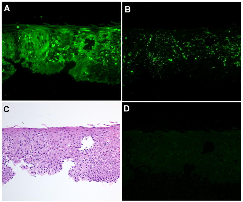
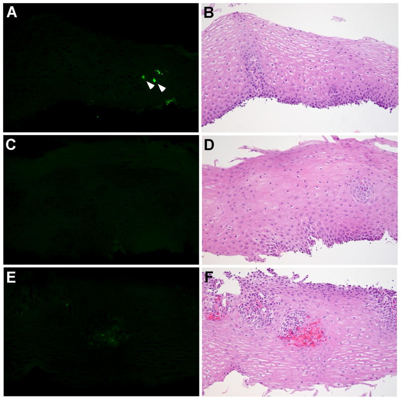
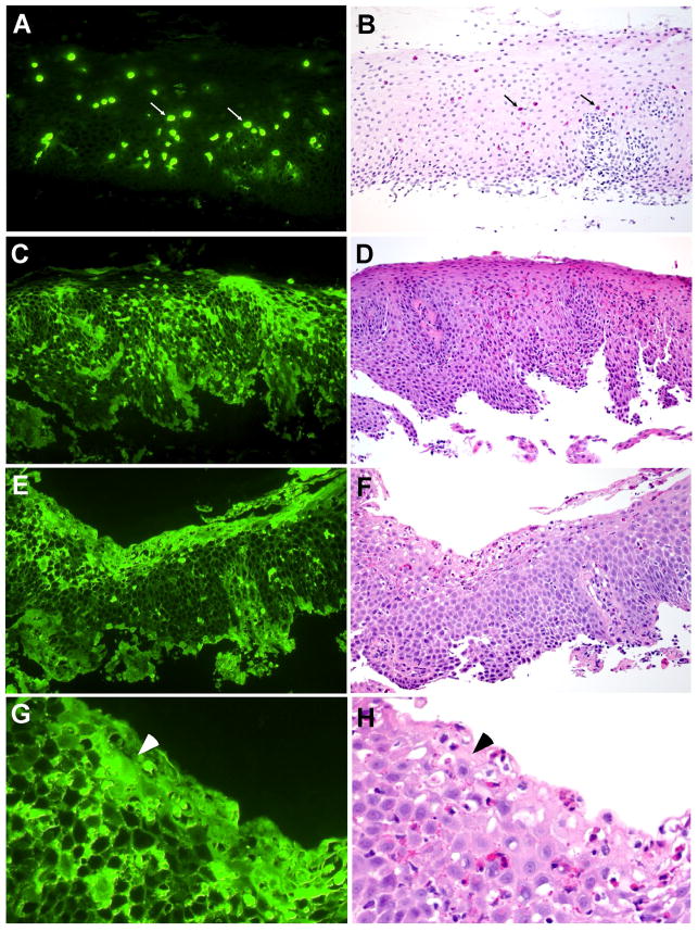
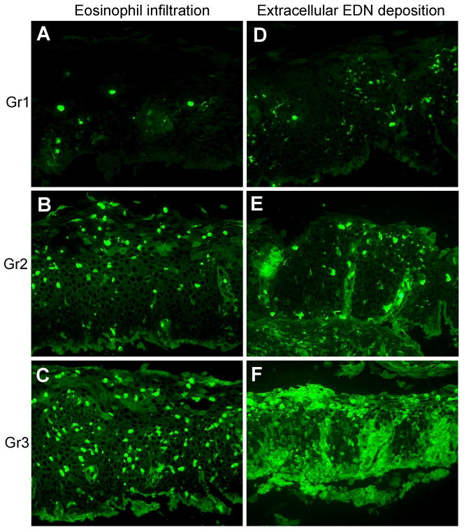
Methods: We studied specimens from 10 adult EoE patients and eight histologically normal controls (three under age 17). Sections from mid-esophageal biopsy specimens were stained for EDN by immunofluorescence, using a polyclonal rabbit antibody to EDN. Cellular staining (i.e., infiltration of intact eosinophils) and extracellular staining (i.e., deposition of released EDN) were scored in a blinded manner on an established 7-point scale.
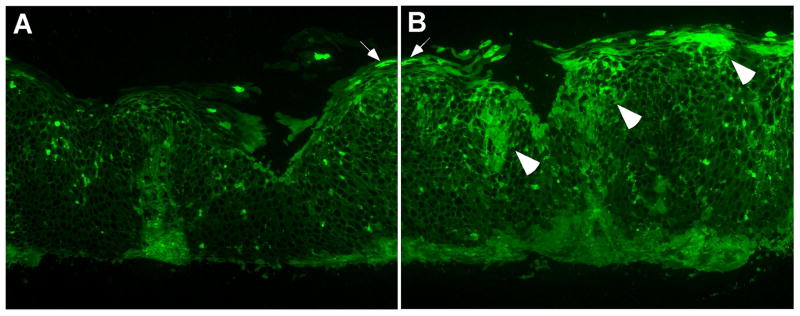
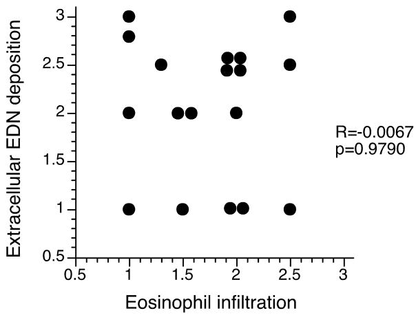
Results: Esophageal biopsy specimens from histologically normal controls showed no or few intact eosinophils and no or minimal extracellular EDN deposition. In contrast, EDN staining was clearly observed in specimens from all EoE patients. In some EoE patients, marked extracellular EDN deposition was observed despite relatively small numbers of intact eosinophils. Overall, there was no correlation between the eosinophil infiltration and the extracellular EDN staining scores.
Conclusions: Marked tissue deposition of extracellular EDN is present in the esophagus of EoE patients. Tissue eosinophil counts may underestimate how extensively eosinophils are involved, particularly in individuals with marked eosinophil degranulation. Evaluation of EDN staining in esophageal biopsy specimens may be useful to diagnose and manage patients with EoE.
Conflict of interest statement
Figures
References
-
- Attwood SEA, Smyrk TC, Demeester TR, et al. Esophageal eosinophilia with dysphagia: A distinct clinicopathologic syndrome. Dig Dis Sci. 1993;38:109–16. - PubMed
-
- Furuta GT, Liacouras CA, Collins MH, et al. Eosinophilic esophagitis in children and adults: A systematic review and consensus recommendations for diagnosis and treatment. Gastroenterology. 2007;133:1342–63. - PubMed
-
- Blanchard C, Wang N, Rothenberg ME. Eosinophilic esophagitis: Pathogenesis, genetics, and therapy. J Allergy Clin Immunol. 2006;118:1054–59. - PubMed
-
- Collins MH. Histopathologic features of eosinophilic esophagitis. Gastrointest Endoscopy Clin N Am. 2008;18:59–71. - PubMed
-
- Chang F, Anderson S. Clinical and pathological features of eosinophilic oesophagitis: a review. Pathology. 2008;40:3–8. - PubMed
Publication types
MeSH terms
Substances
Grants and funding
LinkOut - more resources
Full Text Sources
Other Literature Sources
Medical
