. 2010 Apr;18(4):421-8.
doi: 10.1038/ejhg.2009.186. Epub 2009 Nov 4.

Copy number variation upstream of PMP22 in Charcot-Marie-Tooth disease

Affiliations

Copy number variation upstream of PMP22 in Charcot-Marie-Tooth disease

Marian A J Weterman et al. Eur J Hum Genet. 2010 Apr.

Abstract

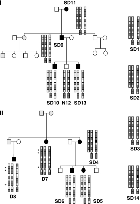
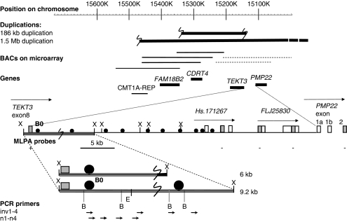
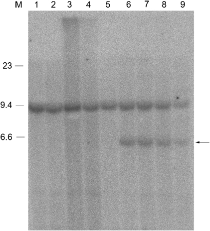
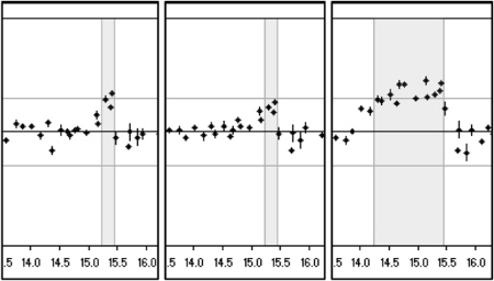
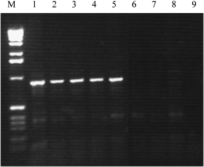
In several individuals with a Charcot-Marie-Tooth (CMT) phenotype, we found a copy number variation (CNV) on chromosome 17p12 in the direct vicinity of the peripheral myelin protein 22 (PMP22) gene. The exact borders and size of this CNV were determined by Southern blot analysis, MLPA, vectorette PCR, and microarray hybridization analyses. All patients from six apparently unrelated families carried an identical 186-kb duplication different from the commonly reported 1.5-Mb duplication associated with CMT1A. This ancestral mutation that was not reported in the human structural variation database was only detected in affected individuals and family members. It was absent in 2124 control chromosomes and 40 patients with a chronic inflammatory demyelinating polyneuropathy (CIDP) and therefore should be regarded as causative for the disease. This variant escapes most routine diagnostic screens for CMT1A, because copy numbers of PMP22 probes were all normal. No indications were found for the involvement of the genes that are located within this duplication. A possible association of this duplication with a mutation in the PMP22 coding regions was also excluded. We suggest that this CNV proximal of the PMP22 gene leads to CMT through an unknown mechanism affecting PMP22 expression.


Figures

Figure 1
Figure 1
Pedigrees of Charcot–Marie–Tooth (CMT) families I and II and haplotype of patients. Family trees of both families are depicted with affected members with filled symbols. Gray symbols indicate that the phenotype could not be certainly assessed or was not known. All affected members and unaffected member N12 from family I were screened for the presence of duplications. Patients D7 and D8 from family II carried the conventional 1.5-Mb duplication instead of the 186-kb duplication present in patients SD4–6. TEKT3 and PMP22 polymorphisms are represented as squares in the order as they appear on the coding/-strand of the chromosome from centromere to telomere (nine TEKT3 and two PMP22 SNPs). For reasons of clarity the (inferred) haplotypes of the tandem duplication are depicted next to each other. Dark shaded squares represent the most frequently occurring allele, and the light shaded squares the minor allele. In one case, for rs11411664, frequencies were unknown and the alleles were represented by black and white boxes, respectively. Represented SNPs are rs396445, rs7226363, rs2305959, rs230901, rs11411664, rs230898, rs230897, rs2286516, and rs13961 of the TEKT3 gene and rs231020 and rs3744333 of PMP22. Known frequencies of the TEKT3 alleles associated with the small duplication of represented SNPs are 0.14–0.18, 0.135–0.217, 0.2, 0.9, 0.47–0.54, 0.475, 0.7, 0.217–0.25, respectively. The haplotypes of single cases SD1–3 and SD14 are also provided in the same manner.
Figure 2
Figure 2
Schematic overview of junction region: genes and duplicated regions on chromosome 17. The 1.5-Mb duplication is only partially drawn as indicated by the dotted line on the end because it is larger than the depicted region. BACs with a duplicated signal in the microarrays are represented by a solid thick line, the BAC with a normal signal by a thin line and the two BACs that showed partial duplication by a dotted line. The region between TEKT3 and PMP22 is shown in detail; E, B, and X represent EcoRI, BamH1, and XbaI restriction sites, filled circles represent probes used for Southern blot analysis, gray squares represent the positions of the MLPA probes. The normal 9.2-kb XbaI fragment and aberrant 6-kb XbaI fragment that were detected by Southern blot analysis by probe Bo and contain the location of the junction (large S), are depicted at the bottom. Vectorette PCR primed with inv1 or n2 and nested primers n1–n4 gave expected products of 3–4 kb in size all containing the duplication junction.
Figure 3
Figure 3
Southern blot analysis reveals the junction fragment. Southern blot analysis using B0 as a probe and XbaI-digested DNA from patients with the 1.5-Mb duplication (lanes 1, 3) without duplication from family I (lane 2) or from unrelated individuals (lanes 4 and 5), and from patients with the smaller duplication (lanes 6–9). In addition to the normal XbaI fragment of 9.2 kb that is detected with this probe, a junction fragment can also be seen of approximately 6 kb in patients carrying the smaller duplication (arrow). On the left, three bands of the lambda-HindIII marker are shown.
Figure 4
Figure 4
Microarray analysis of duplicated region. Microarray CGH analysis of the same region on chromosome 17 from 17p13.3 to 17p11.2 (position in Mb on X-axis; ratio on Y-axis; upper horizontal line depicts the 1.2 cutoff) for two patients with the small duplication (left two panels) and one with the conventional 1.5-Mb duplication (right panel). The duplicated region (shaded) clearly shows three duplicated BAC signals in the middle of this box for the two patients with the small duplication (RP11-726O12, RP11-378O18, RP11-765E8). The two BACs on the distal side (RP11-686G16, RP11-655L10) still show a partial duplication (see also dotted lines in Figure 2).
Figure 5
Figure 5
Detection of genomic junction fragments. PCR performed directly on genomic DNA from five patients with the 186-kb duplication (lanes 1–5), a patient with the 1.5-Mb deletion (lane 6) or 1.5-Mb duplication (lane 7) or on normal DNA (lane 8) using primers located at 258 and 593 bp from the opposite sites of the junction breakpoints, respectively. Lane 9 contains the water PCR control. The molecular marker shown is the 1-kb ladder (Invitrogen Life Science, Breda, The Netherlands).

References

    1. Bailey JA, Gu Z, Clark RA, et al. Recent segmental duplications in the human genome. Science. 2002;297:1003–1007. - PubMed
    1. Chance PF, Alderson MK, Leppig KA, et al. DNA deletion associated with hereditary neuropathy with liability to pressure palsies. Cell. 1993;72:143–151. - PubMed
    1. Lupski JR, Oca-Luna RM, Slaugenhaupt S, et al. DNA duplication associated with Charcot-Marie-Tooth disease type 1A. Cell. 1991;66:219–232. - PubMed
    1. Pentao L, Wise CA, Chinault AC, Patel PI, Lupski JR. Charcot-Marie-Tooth type 1A duplication appears to arise from recombination at repeat sequences flanking the 1.5 Mb monomer unit. Nat Genet. 1992;2:292–300. - PubMed
    1. Matsunami N, Smith B, Ballard L, et al. Peripheral myelin protein-22 gene maps in the duplication in chromosome 17p11.2 associated with Charcot-Marie-Tooth 1A. Nat Genet. 1992;1:176–179. - PubMed

Publication types

MeSH terms