Review
. 2010 Jul 1;13(1):39-75.
doi: 10.1089/ars.2009.2733.

Oxidized LDL: diversity, patterns of recognition, and pathophysiology

Affiliations
Review

Oxidized LDL: diversity, patterns of recognition, and pathophysiology

Irena Levitan et al. Antioxid Redox Signal. .

Abstract

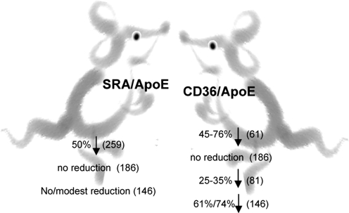
Oxidative modification of LDL is known to elicit an array of pro-atherogenic responses, but it is generally underappreciated that oxidized LDL (OxLDL) exists in multiple forms, characterized by different degrees of oxidation and different mixtures of bioactive components. The variable effects of OxLDL reported in the literature can be attributed in large part to the heterogeneous nature of the preparations employed. In this review, we first describe the various subclasses and molecular composition of OxLDL, including the variety of minimally modified LDL preparations. We then describe multiple receptors that recognize various species of OxLDL and discuss the mechanisms responsible for the recognition by specific receptors. Furthermore, we discuss the contentious issues such as the nature of OxLDL in vivo and the physiological oxidizing agents, whether oxidation of LDL is a prerequisite for atherogenesis, whether OxLDL is the major source of lipids in foam cells, whether in some cases it actually induces cholesterol depletion, and finally the Janus-like nature of OxLDL in having both pro- and anti-inflammatory effects. Lastly, we extend our review to discuss the role of LDL oxidation in diseases other than atherosclerosis, including diabetes mellitus, and several autoimmune diseases, such as lupus erythematosus, anti-phospholipid syndrome, and rheumatoid arthritis.


Figures

FIG. 1.
FIG. 1.
Structures of PAPC (16:0-20:4 PC) and selected oxidation products. See text for abbreviations.
FIG. 2.
FIG. 2.
Potential pathways of MM-LDL formation in vivo. The physiological modification of LDL takes place by a variety of reactions, both enzymatic and nonenzymatic. The products of all these reactions can be rightfully designated as MM-LDL, although the oxidation may not be the primary event in many of these modifications. Lipid peroxidation is the primary reaction only in the lipoxygenase and free radical mediated pathways. Hydrolysis of SM by secretory SMase C may occur in acute phase response when the SMase level is increased in plasma (305) or by the putative SMase intrinsic to LDL (103). The hydrolysis of LDL SM to ceramide increases the oxidative susceptibility of LDL (271), and also results in the formation of aggregated LDL (249) that is superior to OxLDL in loading of macrophages with cholesterol. The action of sPLA2 on LDL produces an LPC-enriched LDL that should have strong chemotactic and pro-inflammatory effects. PAF acetylhydrolase (Lp-PLA2) may be responsible for the hydrolysis of oxidatively truncated PC in LDL, releasing the cytotoxic aldehydes in addition to LPC. Desialylated LDL has been shown to be present in circulation (279), and is formed either by the action of sialidase or by free radical mediated reactions. This LDL was shown to promote foam cell formation. Glycation of LDL, which is more prevalent in diabetes (80), also increases foam cell formation, and increases the susceptibility of LDL to oxidation. The myeloperoxidase (MPO)-mediated oxidation of LDL results primarily in the modification of tyrosine residues of Apo B (97).
FIG. 3.
FIG. 3.
Mechanisms of OxLDL recognition by different scavenger receptors. Multiple types of scavenger receptors have been identified to recognize and interact with different forms of OxLDL. The major scavenger receptors responsible for OxLDL uptake by macrophages (MF) are: class A scavenger receptors SRAI/II and class B scavenger receptor CD36. OxLDL immune complexes OxLDL are recognized and metabolized via Fcγ receptors. OxLDL, particularly MM-LDL may also be recognized by TLR-4 receptors. Each of these receptors recognizes a different component of the OxLDL particle with SRAI/II receptors recognizing modification of the Apo B protein, CD36 recognizing oxidized phospholipids, and TLR-4 recognizing oxidized cholesteryl esters. The major OxLDL uptake pathway in endothelial cells (ECs) is LOX-1 receptor that also recognizes Apo B modifications. ECs also express CD36 and other types of scavenger receptors. Scavenger receptors are also expressed in other cell types, including smooth muscle cells and platelets. Receptor structures represent the basic domain architecture of the different receptors [receptor structures are adapted from (224)].
FIG. 4.
FIG. 4.
Structural modification of Apo B lysine residues critical for OxLDL recognition by SRAI/II receptors [adapted from (85)]. The major modifications of the Apo B protein leading to the recognition by the SRA receptors include oxidation by lipid oxidation products, such as malondialdehyde, hydroxynoneanl, or trancated phospholipids, as well as acetylation and succinylation. All the modifications occur on the lysine residues with 15%–60% of lysines being required to be modified for the interaction with SRA receptors. The products of these modifications are malondialdehyde, acetic anhydride, and succinic anhydride for oxidation, acetylation, and succinylation, respectively. All three modifications result in lysine charge change with the net change per lysine being Δ = −1 for oxidation and acetylation and Δ = −2 for succinylation.
FIG. 5.
FIG. 5.
The core structural motif of oxidized phospholipids responsible for OxLDL recognition by CD36 receptors [from (228)]. The structural requirements for OxPC-CD36 interaction were identified by OxLDL lipid extraction, fractionation by reverse phase HPLC, and then testing the ability of different lipids to inhibit the binding of NO2-LDL to HEK293 cells transfected with CD36. Molecular structures for the major biologically active constituents were determined by tandem mass spectrometry [for more detail, see (228)]. The figure shows the core structural motif conserved among different various oxidized PC species that support their binding to CD36.
FIG. 6.
FIG. 6.
Structures of cholesteryl ester hydroperoxides responsible for OxLDL–TLR-4 interaction [from (93)]. Cholesteryl ester hydroperoxides have been identified as the biological components of MM-LDL responsible for its interaction with TLR-4 receptors by comparing the lipid profiles of MM-LDL with unmodified LDL and then testing the biological activities of the different components. The figure shows the structure of cholesteryl arachidonate, one of the most common cholesteryl esters found in LDL and the fatty acid portion of cholesteryl arachidonate hydroperoxide (15-HpETE) (R stands for cholesterol) that is responsible for the biological activity of MM-LDL. Similar observations were made for cholesteryl linoleate, another common cholesteryl ester of LDL.
FIG. 7.
FIG. 7.
Summary for the relationship between different scavenger receptors and degree of LDL oxidation.
FIG. 8.
FIG. 8.
Importance of intracellular reactive oxygen species (ROS) in the effects of oxidized LDL. Evidence indicates that a common cellular event following the binding of OxLDL to various membrane receptors is the generation of intracellular ROS. The stimulation of pro-inflammatory genes, apoptotic events, and calcium mobilization in the cells are all preceded by ROS generation through the various mechanisms shown. It should, however, be pointed out that not all reactions shown here take place in every cell type. An important consequence of ROS generation is the ‘feed forward’ effect of further oxidation of LDL by the ROS secreted from the cells. The active molecules in OxLDL responsible for the individual pathways have been identified at least in some cases. (For interpretation of the references to color in this figure legend, the reader is referred to the web version of this article at www.liebertonline.com/ars).
FIG. 9.
FIG. 9.
Decrease in the area of atherosclerotic lesions in SRAI/II/Apo E and CD36/Apo E double KO mice. Targeted disruption of SRAI/II receptors in ApoE KO mice was shown to decrease the area of atherosclerotic lesions in some but not in all studies. Similar observations were made on the other pro-atherogenic genetic backgrounds. Disruption of CD36 on the Apo E-deficient background resulted in the decrease in atherosclerotic lesions in the majority but also not in all studies. Several factors, such as severity of the disease, duration of the diet, and the specific site of the vascular tree that was analyzed in these studies were suggested to be responsible for the variability in the responses. Numbers in parenthesis are references.

References

    1. Acton S. Rigotti A. Landschulz KT. Xu S. Hobbs HH. Krieger M. Identification of scavenger receptor SR-BI as a high density lipoprotein receptor. Science. 1996;271:518–520. - PubMed
    1. Acton SL. Scherer PE. Lodish HF. Krieger M. Expression cloning of SR-BI, a CD36-related class B scavenger receptor. J Biol Chem. 1994;269:21003–21009. - PubMed
    1. Adachi H. Tsujimoto M. Endothelial scavenger receptors. Prog Lipid Res. 2006;45:379–404. - PubMed
    1. Akiba S. Chiba M. Mukaida Y. Sato T. Involvement of reactive oxygen species and SP-1 in fibronectin production by oxidized LDL. Biochem Biophys Res Commun. 2003;310:491–497. - PubMed
    1. Allen LB. Capps BE. Miller EC. Clemmons DR. Maile LA. Glucose-oxidized low-density lipoproteins enhance insulin-like growth factor I-stimulated smooth muscle cell proliferation by inhibiting integrin-associated protein cleavage. Endocrinology. 2009;150:1321–1329. - PMC - PubMed

Publication types

MeSH terms