. 2009 Nov 4:4:46.
doi: 10.1186/1750-1326-4-46.

Expression of SORL1 and a novel SORL1 splice variant in normal and Alzheimers disease brain

Affiliations

Expression of SORL1 and a novel SORL1 splice variant in normal and Alzheimers disease brain

Karrie E Grear et al. Mol Neurodegener. .

Abstract

Background: Variations in sortilin-related receptor (SORL1) expression and function have been implicated in Alzheimers Disease (AD). Here, to gain insights into SORL1, we evaluated SORL1 expression and splicing as a function of AD and AD neuropathology, neural gene expression and a candidate single nucleotide polymorphism (SNP).

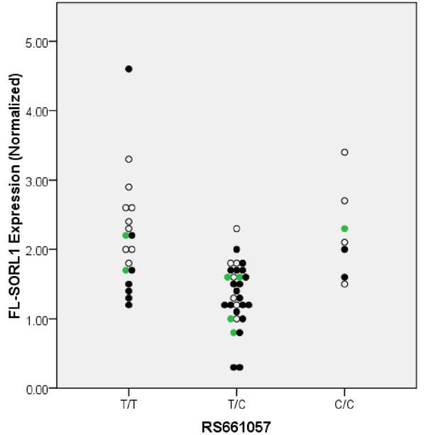
Results: To identify SORL1 splice variants, we scanned each of the 46 internal SORL1 exons in human brain RNA samples and readily found SORL1 isoforms that lack exon 2 or exon 19. Quantification in a case-control series of the more abundant isoform lacking exon 2 (delta-2-SORL1), as well as the "full-length" SORL1 (FL-SORL1) isoform containing exon 2 showed that expression of FL-SORL1 was reduced in AD individuals. Moreover, FL-SORL1 was reduced in cognitively intact individuals with significant AD-like neuropathology. In contrast, the expression of the delta-2-SORL1 isoform was similar in AD and non-AD brains. The expression of FL-SORL1 was significantly associated with synaptophysin expression while delta-2-SORL1 was modestly enriched in white matter. Lastly, FL-SORL1 expression was associated with rs661057, a SORL1 intron one SNP that has been associated with AD risk. A linear regression analysis found that rs661057, synaptophysin expression and AD neuropathology were each associated with FL-SORL1 expression.

Conclusion: These results confirm that FL-SORL1 expression declines in AD and with AD-associated neuropathology, suggest that FL-SORL1 declines in cognitively-intact individuals with AD-associated neuropathology, identify a novel SORL1 splice variant that is expressed similarly in AD and non-AD individuals, and provide evidence that an AD-associated SNP is associated with SORL1 expression. Overall, these results contribute to our understanding of SORL1 expression in the human brain.


Figures

Figure 1
Figure 1
Evaluation of SORL1 Exonic Splicing. Inefficiently spliced exons were detected by amplifying between the indicated exons. The novel splice variants that were identified and confirmed by sequencing are marked with asterisks. Since these representative results are a montage of multiple experiments, the molecular weight markers are approximate.
Figure 2
Figure 2
Sequence of SORL1 Isoforms. The nucleotide and protein sequence of FL-SORL1 as well as sequences corresponding to each of the novel isoforms is shown. Figure 2A (upper panel) depicts FL-SORL1 nucleotide and protein sequence beginning with the ATG translation start site in exon 1 through exon 3. Exon 2 is marked in red font. The nucleotide and protein sequence corresponding to delta-2-SORL1, i.e., loss of amino acids V96-D134, is shown in the lower panel, with the exon 1-3 junction indicated by red font. Figure 2B shows FL-SORL1 nucleotide and protein sequence from exon 18-20 (upper panel) with exon 19 marked in red font. The lower panel shows the nucleotide and protein sequence corresponding to delta-19-SORL1, i.e., a novel peptide sequence encoded by an out-of-frame exon 20, followed by a premature stop codon. The exon 18-20 junction is indicated by red font and the novel peptide sequence is shown in blue font.
Figure 3
Figure 3
Quantitation of FL-SORL1 and Delta-2-SORL1 in AD and non-AD Brain. Real time PCR quantification of each isoform shows that FL-SORL1 and delta-2-SORL1 vary in parallel (A) (r2 = 0.371, p < 0.001, AD individuals represented as filled circles, non-AD as open circles). FL-SORL1 was decreased in AD relative to non-AD individuals (B) while delta-2-SORL1 was similar in AD and non-AD individuals (C). Delta-2-SORL1 as a percentage of total SORL1 expression was increased in AD individuals (D).
Figure 4
Figure 4
Quantitation of FL-SORL1 and Delta-2-SORL1 as a Function of Braak Stage. The expression of FL-SORL1 was reduced in cognitively intact individuals with a Braak stage indicative of some AD neuropathology relative to cognitively intact individuals with little neuropathology (A). In contrast, the expression of delta-2-SORL1 was similar among the groups (B).
Figure 5
Figure 5
Quantitation of FL-SORL1 as a function of synaptophysin expression and Braak stage. Individuals that have Braak stages that are low (0-II, open circles), moderate (Braak III-V, green circles) or high (VI, black-filled circles) are indicated. Linear regression analyses found that both synaptophysin and Braak stage (two degrees of freedom) were significantly associated with SORL1 expression, (p < 0.001 and p = 0.015, respectively). Synaptophysin expression showed only a slight trend towards decreased expression with Braak stage, which did not achieve significance (p = 0.55, ANOVA)
Figure 6
Figure 6
Quantitation of FL-SORL1 as a function of rs661057 and Braak stage. Individuals that have Braak stages that are low (0-II, open circles), moderate (Braak III-V, green circles) or high (VI, black-filled circles) are indicated.

References

    1. Jacobsen L, Madsen P, Jacobsen C, Nielsen MS, Gliemann J, Petersen CM. Activation and functional characterization of the mosaic receptor SorLA/LR11. J Biol Chem. 2001;276:22788–96. doi: 10.1074/jbc.M100857200. - DOI - PubMed
    1. Andersen OM, Reiche J, Schmidt V, Gotthardt M, Spoelgen R, Behlke J, von Arnim CA, Breiderhoff T, Jansen P, Wu X, Bales KR, Cappai R, Masters CL, Gliemann J, Mufson EJ, Hyman BT, Paul SM, Nykjaer A, Willnow TE. Neuronal sorting protein-related receptor sorLA/LR11 regulates processing of the amyloid precursor protein. Proc Natl Acad Sci USA. 2005;102:13461–6. doi: 10.1073/pnas.0503689102. - DOI - PMC - PubMed
    1. Offe K, Dodson SE, Shoemaker JT, Fritz JJ, Gearing M, Levey AI, Lah JJ. The lipoprotein receptor LR11 regulates amyloid beta production and amyloid precursor protein traffic in endosomal compartments. J Neurosci. 2006;26:1596–603. doi: 10.1523/JNEUROSCI.4946-05.2006. - DOI - PMC - PubMed
    1. Dodson SE, Andersen OM, Karmali V, Fritz JJ, Cheng D, Peng J, Levey AI, Willnow TE, Lah JJ. Loss of LR11/sorLA enhances early pathology in a mouse model of amyloidosis: Evidence for a proximal role in Alzheimer's disease. J Neurosci. 2008;28:12877–86. doi: 10.1523/JNEUROSCI.4582-08.2008. - DOI - PMC - PubMed
    1. Scherzer CR, Offe K, Gearing M, Rees HD, Fang G, Heilman CJ, Schaller C, Bujo H, Levey AI, Lah JJ. Loss of apolipoprotein E receptor LR11 in Alzheimer disease. Arch Neurol. 2004;61:1200–5. doi: 10.1001/archneur.61.8.1200. - DOI - PubMed

LinkOut - more resources