Review
. 2010 Feb;300(2-3):193-204.
doi: 10.1016/j.ijmm.2009.10.003. Epub 2009 Nov 3.

Interaction of staphylococci with bone

Affiliations
Review

Interaction of staphylococci with bone

John A Wright et al. Int J Med Microbiol. 2010 Feb.

Abstract

Staphylococci, in particular Staphylococcus aureus, are the predominant cause of bone infections worldwide. These infections are painful, debilitating and with the rise in antibiotic-resistant forms, increasingly difficult to treat. The growth in the number of prosthetic joint replacement procedures also provides new opportunities for these infections to take hold. Comprehending the mechanisms by which staphylococci interact with and damage bone is critical to the development of new approaches to meet this challenge. This review summarises current understanding of the mechanisms by which staphylococci infect and damage bone. We address the role of the inflammatory response to staphylococcal infection in disrupting the homeostatic balance of bone matrix deposition and resorption and thereby mediating bone destruction. A number of virulence factors that have been shown to contribute to bone infection and pathology are discussed, however no single factor has been defined as being specific to bone infections. Although traditionally considered an extracellular pathogen, there is increasing evidence that staphylococci are able to invade host cells, and that an intracellular lifestyle may facilitate long-term persistence in bone tissue, enabling evasion of antimicrobials and host immune responses. 'Small colony variant' strains, with mutations disabling the electron transport pathway appear particularly adept at invading and persisting within host cells, and exhibit enhanced antimicrobial resistance, and may represent a further complication in the treatment and management of staphylococcal bone disease.


Figures

Fig. 1
Fig. 1
Schematic diagram of IL-1 and TNF signal transduction events. (A) IL-1 binding to IL-1RI results in recruitment of the IL-1 receptor accessory protein IL-1RAcP, which form a complex that, via the intracellular Toll/IL-1 receptor (TIR) domains of the two receptor molecules, engages the MyD88 (myeloid differentiation factor 88) adaptor protein (Arend et al., 2008; Dinarello, 2009; Verstrepen et al., 2008). MyD88 binding initiates phosphorylation of IRAK-4 (interleukin-1 receptor-associated kinase 4), IRAK-2 and IRAK-1. IRAK-1 recruits TRAF6 and these two proteins localise to the cell membrane where they associate with TAK1 (TGF-β-activated kinase 1), TAB1 (TAK1-binding protein) and TAB2. The TAK1/TAB1/TAP2/TRAF6 complex translocates to the cytosol where TRAF-6 is poly-ubiquitinated and TAK1 is subsequently phosphorylated, and activates NF-κB, p38 MAPK and JNK. The MyD88 adaptor molecule and downstream signalling events are common to most Toll-like receptors (TLRs), and, via TRAF6, this pathway also converges with RANKL signalling. (B) Engagement of the homotrimeric TNF-R1 receptor by TNF enables recruitment of TRADD (TNF-R1 associated death domain protein) via homotypic death domain interactions. TRADD associates with itself, TRAF2 and RIP1 (receptor-interacting protein 1) and with TRAF5. TNF-R2 is able to directly associate with TRAF2, independently of TRADD. TRAF2 auto-poly-ubiquitinates and ubiquitinates RIP1. Poly-ubiquitination of RIP1 then leads to the recruitment of TAK1, through interactions with TAB1, TAB2 and TAB3, again leading to NF-κB, p38 MAPK and JNK activation (Bradley, 2008; Verstrepen et al., 2008).
Fig. 2
Fig. 2
Schematic diagram of IL-6 signalling events. IL-6 specifically binds to IL-6 receptor α (IL-6Rα), and recruits a homodimer of the gp130 signal-transducing protein (Naugler and Karin, 2008). This in turn leads to association of the JAK kinases JAK1, JAK2 and TYK2 with gp130, and the autophosphorylation of these kinases (Heinrich et al., 2003). JAK1 is of particular importance in IL-6 signalling, and cells deficient in this kinase exhibit substantial signalling impairment (Guschin et al., 1995; Heinrich et al., 2001; Rodig et al., 1998). Following JAK activation, gp130 is phosphorylated on multiple tyrosine residues, enabling recruitment and phosphorylation of STAT family transcripton factors. STAT3 is the major transcription factor acting downstream of IL-6 signalling, and dimerises upon phosphorylation and translocates to the nucleus to activate transcription of an array of target genes (Li et al., 2002; Naugler and Karin, 2008). IL-6 also activates the MAPK pathway, via JAK phosphorylation of SHP2 (SH2 domain-containing protein-tyrosine phosphatase). SHP2 is thought to recruit either the Grb2-SOS (growth factor receptor-bound protein/Son of Sevenless) complex and/or Gab1 (Grb2-associated binder 1) to gp130. Recruitment of SOS then enables activation of Ras and subsequently the MAPK (mitogen-associated protein kinase) pathway, although the importance of this pathway is currently unclear (Heinrich et al., 2003; Naugler and Karin, 2008).
Fig. 3
Fig. 3
Schematic diagram of some of the signalling events involved in invasion of ‘non-professional phagocytic’ host cells by S. aureus. FnBPs expressed on the bacterial surface bind to fibronectin molecules in the extracellular matrix, which form a bridge with integrin α5β1 on the host cell membrane (Dziewanowska et al., 1999; Fowler et al., 2000; Sinha et al., 1999). Ligation of this complex to the integrin receptor causes receptor clustering and subsequent activation of ILK, via interaction with the β1 subunit (Wang et al., 2006). There is subsequent recruitment of paxillin and FAK and downstream phosphorylation of cortactin leading to remodelling of the actin cytoskeleton to enable bacterial uptake (Agerer et al., 2005). The focal adhesion proteins zyxin, vinculin and tensin are also recruited to the site of bacterial uptake, the latter two potentially via interactions with paxillin and FAK, respectively (Agerer et al., 2005).

References

    1. Abdelnour A., Arvidson S., Bremell T., Ryden C., Tarkowski A. The accessory gene regulator (agr) controls Staphylococcus aureus virulence in a murine arthritis model. Infect. Immun. 1993;61:3879–3885. - PMC - PubMed
    1. Abdelnour A., Bremell T., Tarkowski A. Toxic shock syndrome toxin 1 contributes to the arthritogenicity of Staphylococcus aureus. J. Infect. Dis. 1994;170:94–99. - PubMed
    1. Adler H., Widmer A., Frei R. Emergence of a teicoplanin-resistant small colony variant of Staphylococcus epidermidis during vancomycin therapy. Eur. J. Clin. Microbiol. Infect. Dis. 2003;22:746–748. - PubMed
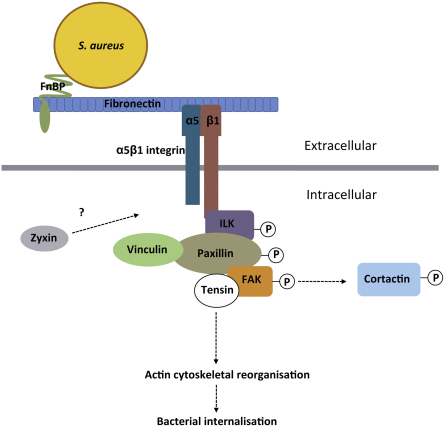
    1. Agerer F., Lux S., Michel A., Rohde M., Ohlsen K., Hauck C.R. Cellular invasion by Staphylococcus aureus reveals a functional link between focal adhesion kinase and cortactin in integrin-mediated internalisation. J. Cell Sci. 2005;118:2189–2200. - PubMed
    1. Aggarwal B.B. Signalling pathways of the TNF superfamily: a double-edged sword. Nat. Rev. Immunol. 2003;3:745–756. - PubMed

Publication types

MeSH terms