. 2010 Jan 8;285(2):1066-74.
doi: 10.1074/jbc.M109.044321. Epub 2009 Nov 4.

Inherent anti-amyloidogenic activity of human immunoglobulin gamma heavy chains

Affiliations

Inherent anti-amyloidogenic activity of human immunoglobulin gamma heavy chains

Sharad P Adekar et al. J Biol Chem. .

Abstract

We have previously shown that a subpopulation of naturally occurring human IgGs were cross-reactive against conformational epitopes on pathologic aggregates of Abeta, a peptide that forms amyloid fibrils in the brains of patients with Alzheimer disease, inhibited amyloid fibril growth, and dissociated amyloid in vivo. Here, we describe similar anti-amyloidogenic activity that is a general property of free human Ig gamma heavy chains. A gamma(1) heavy chain, F1, had nanomolar binding to an amyloid fibril-related conformational epitope on synthetic oligomers and fibrils as well as on amyloid-laden tissue sections. F1 did not bind to native Abeta monomers, further indicating the conformational nature of its binding site. The inherent anti-amyloidogenic activity of Ig gamma heavy chains was demonstrated by nanomolar amyloid fibril and oligomer binding by polyclonal and monoclonal human heavy chains that were isolated from inert or weakly reactive antibodies. Most importantly, the F1 heavy chain prevented in vitro fibril growth and reduced in vivo soluble Abeta oligomer-induced impairment of rodent hippocampal long term potentiation, a cellular mechanism of learning and memory. These findings demonstrate that free human Ig gamma heavy chains comprise a novel class of molecules for developing potential therapeutics for Alzheimer disease and other amyloid disorders. Moreover, establishing the molecular basis for heavy chain-amyloidogenic conformer interactions should advance understanding on the types of interactions that these pathologic assemblies have with biological molecules.


Figures

FIGURE 1.
FIGURE 1.
Fibril and Aβ conformer binding by HC F1 and mAb F2. A and B, binding curves for F1 and F2, respectively, against plate-immobilized amyloid fibrils: LC (○) and Aβ40 (●) fibrils, non-amyloid elastin fibrils (□), and CAPS (▵). C and D, F1 and a N-terminal Aβ-reactive mAb, MAB1560, respectively, binding to plate-immobilized Aβ40 monomer in the presence (○) or absence (●) of a 100-fold molar excess of homologous peptide conformer. Binding studies were carried out in PBSA containing 1% BSA at 37 °C.
FIGURE 2.
FIGURE 2.
Western blot analysis of Ig chain expression by the F1 hybridoma. The blots show that only human HC was detected for the F1 hybridoma cell lysate. Human HC and LC were detected in cell lysates for control hybridomas expressing IgGκ or IgGλ, respectively. The cell fusion partner for the F1 hybridoma, B5-6T, was negative for HC and LC. The cell lysates were run on a 4–12% BisTris gel under reducing conditions, and Western blots were carried out using HRP-conjugated secondary antibodies specific for human HC γ, LC κ, or λ.
FIGURE 3.
FIGURE 3.
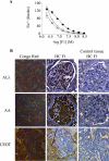
Ex vivo and in situ amyloid fibril binding by HC F1. A, amyloid fibril binding curves for F1 against plate-immobilized fibrils extracted from patients with ALλ6 (○) or AA (▵) amyloidosis or AD (♦). B, representative Congo red and F1 staining of amyloid-laden glomeruli kidney tissue sections from patients with ALλ6 or AA amyloidosis or with an Apin amyloid-containing CEOT tumor (a) that was surrounded by non-amyloid-containing amelobastoma tumor tissue (b). Panels on the right show negative F1 staining of control tissues that did not contain amyloid. The original magnification for tissue sections was ×200.
FIGURE 4.
FIGURE 4.
Aβ fibril-binding by monoclonal and polyclonal IgGs and γ HCs isolated from healthy individuals. A and B, Aβ40 fibril-binding curves for mAbs 13A and 30B, respectively, for whole molecule (♦) and HC (○), as well as binding by HC F1 (●). C and D, Aβ40 fibril binding by polyclonal HCs derived from five different subjects (●, ○, □, ■, and ▵) and polyclonal IgGs (♦, ◇, and +).
FIGURE 5.
FIGURE 5.
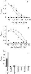
Specificity of HCs 13A and 30B for amyloidogenic conformers. A, Aβ fibril binding for reduced (▵) and carboxymethylated (▴) HC 13A and for the intact IgG (●). B, Aβ fibril binding for reduced (▵) and carboxymethylated (▴) HC 30B and for the intact IgG (●). C, Aβ fibril and non-amyloidogenic molecule binding by 50 nm carboxymethylated 13A and 30B HCs (closed and open bars, respectively). IgG and HC binding was determined in PBSA containing 1% BSA by ELISA using a HRP-streptavidin detection system.
FIGURE 6.
FIGURE 6.
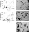
HC F1 inhibition of de novo Aβ fibril growth. A, dose-dependent effect of 5 μm (○), 2.5 μm (●), 1.25 μm (▴), 0.31 μm (▵), and 0 μm (□) F1 on Aβ40 fibril formation. B, Aβ40 fibril formation carried out in the absence (□) or presence of 0.7 μm HC 13A (●), IgG 13A (○), or BSA (▴). C–E, electron micrographs of aggregate products from reactions carried out in the absence of inhibitor (C) or in the presence of 1 μm F1 (D) or IgG 13A (E). Each reaction was carried out 90 μm Aβ40 with or without inhibitor in PBSA containing 30 μm ThT at 37 °C.
FIGURE 7.
FIGURE 7.
HC F1 inhibition of Aβ fibril elongation. A, dose-dependent inhibitory effect of F1 (▴), mAb 13A (●), 13A HC (○), and BSA (▵) on Aβ40 fibril elongation. B–D, electron micrographs of aggregate products from reactions carried out in the absence of inhibitor (B) or in the presence of 1 μm F1 (C) or IgG 13A (D). Fibril extension reactions were carried out with 30 nm biotinyl-Aβ40 with or without inhibitor in PBSA for 3 h at 37 °C.
FIGURE 8.
FIGURE 8.
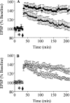
HC F1 prevents Aβ42-mediated inhibition of LTP in the rat hippocampus in vivo. LTP is an activity-dependent long lasting increase in the strength of synaptic connections (EPSPs) and models the cellular mechanisms underlying learning and memory formation. A, in vehicle-injected controls (●), the application of high frequency conditioning stimulation (arrow) at 30 min triggered a robust LTP. Intracerebroventricular injection (asterisk) of synthetic Aβ42 (40 pmol, ▴) 10 min before the conditioning stimulation completely inhibited LTP. B, the inhibition of LTP by Aβ42 was prevented by intracerebroventricular co-injection of 60 pmol (3 μg) of HC F1 (▵). Co-injection of a similar amount (3.75 μg) of the control mAb, 13A (○) did not prevent the Aβ-mediated inhibition of LTP.
FIGURE 9.
FIGURE 9.
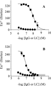
LC binding to Aβ fibrils. Binding curves for LCs (■) and intact IgGs (●) for 13A (A) and 30B (B) against plate-immobilized Aβ fibrils. Antibody binding was determined in PBSA containing 1% BSA by ELISA using a -streptavidin detection system.

References

    1. Stefani M. (2004) Biochim. Biophys. Acta 1739, 5–25 - PubMed
    1. Goedert M., Spillantini M. G. (2006) Science 314, 777–781 - PubMed
    1. Haass C., Schlossmacher M. G., Hung A. Y., Vigo-Pelfrey C., Mellon A., Ostaszewski B. L., Lieberburg I., Koo E. H., Schenk D., Teplow D. B., Selkoe D. J. (1992) Nature 359, 322–325 - PubMed
    1. Hardy J., Selkoe D. J. (2002) Science 297, 353–356 - PubMed
    1. Walsh D. M., Selkoe D. J. (2007) J. Neurochem. 101, 1172–1184 - PubMed

Publication types

LinkOut - more resources