. 2010 Jul;20(7):1685-95.
doi: 10.1093/cercor/bhp232. Epub 2009 Nov 4.

Reduction of basal forebrain cholinergic system parallels cognitive impairment in patients at high risk of developing Alzheimer's disease

Affiliations

Reduction of basal forebrain cholinergic system parallels cognitive impairment in patients at high risk of developing Alzheimer's disease

Michel Grothe et al. Cereb Cortex. 2010 Jul.

Abstract

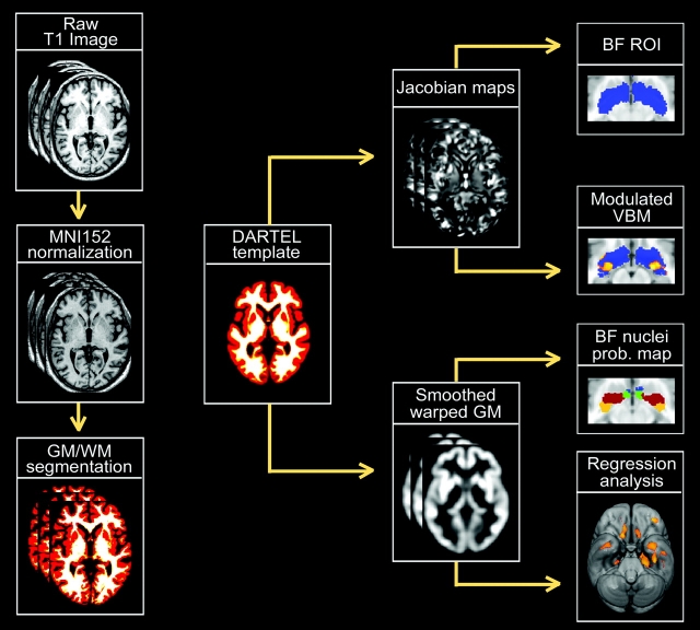
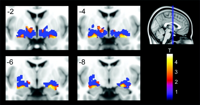
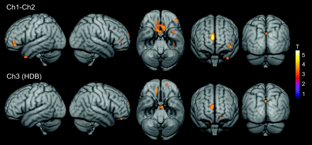
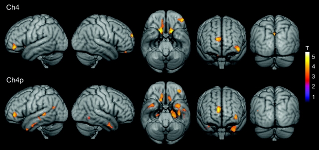
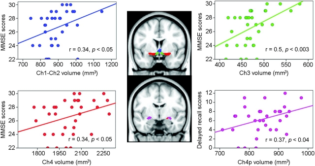
Neuropathological studies suggest that the basal forebrain cholinergic system (BFCS) is affected in Alzheimer's disease (AD), but there is no in vivo evidence of early damage to this system in subjects at high risk of developing AD. Here, we found that mild cognitive impairment (MCI) patients exhibited significant volume reduction of the nucleus basalis of Meynert (NbM) using recently developed probabilistic maps of the BFCS space. In addition, volumes of different magnocellular compartments varied significantly with regional gray matter atrophy in regions known to be affected by AD and were found to correlate with cognitive decline in MCI patients. Bilateral reductions of the horizontal nucleus of the diagonal band of Broca (Ch3) and frontal lobe (medial frontal, orbital, subcallosal gyrus, anterior cingulate, and middle frontal gyrus) were significantly associated with a global decline in cognitive status, whereas volume reduction of the posterior compartment of Ch4 (NbM) and temporal lobe (including hippocampus, entorhinal cortex, and amygdala) were associated with impaired delayed recall in MCI patients. These findings establish, for the first time, a link between degeneration of specific cholinergic compartments of the BFCS and cognitive-related deficits in subjects at high risk of developing AD.


Figures

Figure 1.
Figure 1.
Flowchart displaying a scheme of the analysis protocol. Left column (starting from top) shows the SPM5 preprocessing steps applied to each individual raw T1 MRI scan. After linearly normalizing each image to the MNI152 space, they were partitioned into different tissue classes: gray matter (GM), white matter (WM), and cerebrospinal fluid. GM and WM maps were then registered to their average by using an efficient large deformation diffeomorphic framework (DARTEL) to minimize anatomical variations between subjects (Ashburner 2007). Jacobian determinant maps were derived from the DARTEL flow fields, log transformed, and analyzed to determine volumetric changes within the BF magnocellular compartments between healthy elderly and MCI patients. The BF region of interest was based on probabilistic maps of BF compartments obtained from postmortem brains (Zaborzsky et al. 2008). In addition, the volume for each BF magnocellular group was obtained by summing up the jacobian determinant values within a mask of each corresponding compartment, which was obtained by applying a threshold above 50% to each probability map. These volumes were then regressed on the smoothed modulated warped gray matter maps.
Figure 2.
Figure 2.
Bilateral volume reductions of BF in MCI patients compared with healthy elderly subjects, no age corrections (P < 0.01, uncorrected). Coronal slices are from anterior to posterior (y = −2 to y = −8) in intervals of 2 mm. Blue area represents the combined probability maps (Ch1−Ch2, Ch3 = HDB, Ch4, and Ch4p) in MNI space including all voxels showing any probability of belonging to a cholinergic nucleus (BF ROI). Significant volume reductions are coded by the color scale (significance levels represented by T-values).
Figure 3.
Figure 3.
Changes in the volume of Ch1−Ch2 (top panel) and Ch3 = HDB (bottom panel) associated with reductions in regional gray matter concentration in MCI patients. Statistical threshold was set at P < 0.005, uncorrected, and cluster extension was set at 50 contiguous voxels as minimum. Significant correlations are coded by the color scale (significance levels represented by T-values).
Figure 4.
Figure 4.
Changes in the volume of Ch4 (top panel) and Ch4p (bottom panel) associated with reductions in regional gray matter concentration in MCI patients. Statistical threshold was set at P < 0.005, uncorrected, and cluster extension was set at 50 contiguous voxel as minimum. Significant correlations are coded by the color scale (significance levels represented by T-values).
Figure 5.
Figure 5.
Scatter plots showing significant relationships between cognitive impairment and volume loss of each BF cholinergic compartment in MCI patients. Note that cognitive function is differently associated with volume reductions of BF nuclei in MCI patients: Global cognitive decline (MMSE scores) are related to volumetric changes in Ch1−Ch2, Ch3 = HDB, and Ch4, whereas impaired delayed recall is associated with volume loss in Ch4p. R (Pearson correlation coefficient) and P (level of significance) values were added to each subplot. Cholinergic nucleus masks are projected onto the MNI152 anatomical space, and color-coded: Ch1–2 (blue), Ch3 = HDB (green), Ch4 (red), and Ch4p (purple). Identical color identification is maintained in scatter plots for each BF nucleus.

References

    1. Amunts K, Schleicher A, Zilles K. Cytoarchitecture of the cerebral cortex– more than localization. Neuroimage. 2007;37:1061–1065. - PubMed
    1. Amunts K, Zilles K. Atlases of the human brain: tools for functional neuroimaging. In: Zaborsky L, Wouterlood FG, Lanciego JL, editors. Neuroanatomical tract-tracing 3: molecules, neurons, and systems. 3rd ed. New York: Springer; 2006. pp. 566–603.
    1. Arendt T, Bigl V, Tennstedt A, Arendt A. Neuronal loss in different parts of the nucleus basalis is related to neuritic plaque formation in cortical target areas in Alzheimer's disease. Neuroscience. 1985;14:1–14. - PubMed
    1. Artero S, Ancelin ML, Portet F, Dupuy A, Berr C, Dartigues JF, Tzourio C, Rouaud O, Poncet M, Pasquier F, et al. Risk profiles for mild cognitive impairment and progression to dementia are gender specific. J Neurol Neurosurg Psychiatry. 2008;79:979–984. - PubMed
    1. Ashburner J. A fast diffeomorphic image registration algorithm. Neuroimage. 2007;38:95–113. - PubMed

Publication types

MeSH terms