. 2010 Jan;84(2):983-92.
doi: 10.1128/JVI.02039-09. Epub 2009 Nov 4.

Characterization of Lassa virus glycoprotein oligomerization and influence of cholesterol on virus replication

Affiliations

Characterization of Lassa virus glycoprotein oligomerization and influence of cholesterol on virus replication

Katrin Schlie et al. J Virol. 2010 Jan.

Abstract

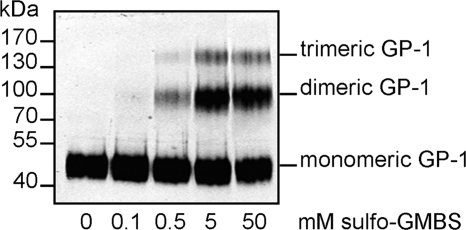
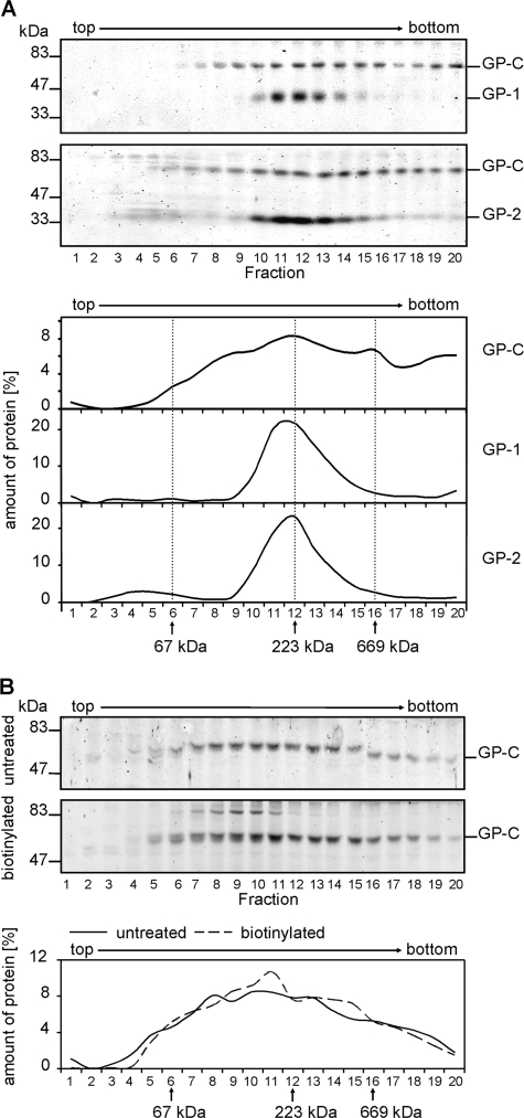
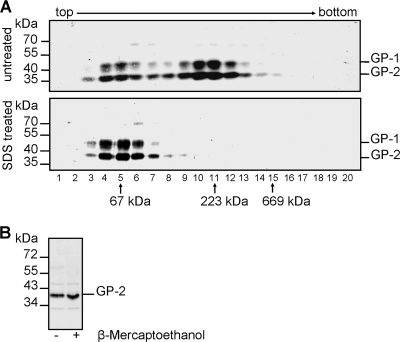
Mature glycoprotein spikes are inserted in the Lassa virus envelope and consist of the distal subunit GP-1, the transmembrane-spanning subunit GP-2, and the signal peptide, which originate from the precursor glycoprotein pre-GP-C by proteolytic processing. In this study, we analyzed the oligomeric structure of the viral surface glycoprotein. Chemical cross-linking studies of mature glycoprotein spikes from purified virus revealed the formation of trimers. Interestingly, sucrose density gradient analysis of cellularly expressed glycoprotein showed that in contrast to trimeric mature glycoprotein complexes, the noncleaved glycoprotein forms monomers and oligomers spanning a wide size range, indicating that maturation cleavage of GP by the cellular subtilase SKI-1/S1P is critical for formation of the correct oligomeric state. To shed light on a potential relation between cholesterol and GP trimer stability, we performed cholesterol depletion experiments. Although depletion of cholesterol had no effect on trimerization of the glycoprotein spike complex, our studies revealed that the cholesterol content of the viral envelope is important for the infectivity of Lassa virus. Analyses of the distribution of viral proteins in cholesterol-rich detergent-resistant membrane areas showed that Lassa virus buds from membrane areas other than those responsible for impaired infectivity due to cholesterol depletion of lipid rafts. Thus, derivation of the viral envelope from cholesterol-rich membrane areas is not a prerequisite for the impact of cholesterol on virus infectivity.


Figures

FIG. 1.
FIG. 1.
Identification of GP-1 oligomers by chemical cross-linking. Lassa virus virions purified by ultracentrifugation through a sucrose cushion were treated with increasing concentrations of sulfo-GMBS as indicated. The cross-linked proteins were separated by SDS-PAGE on an 8% acrylamide gel and analyzed by immunoblotting. GP was detected using an anti-GP-1 antibody.
FIG. 2.
FIG. 2.
Oligomeric state of LASV glycoprotein expressed in mammalian cells. (A) LASV glycoprotein present in cells analyzed by sucrose gradient ultracentrifugation. LASV glycoprotein-expressing cells were lysed with 1% Triton X-100, and cell lysate was subjected to sucrose gradient ultracentrifugation. Aliquots of each gradient fraction were subjected to SDS-PAGE and immunoblotting using the antibody anti-GP-1 (panel 1) or an anti-GP-2-C antiserum (panel 2). GP-C, GP-1, and GP-2 on the immunoblots were quantified (panels 3 to 5). The positions of the gradient marker proteins bovine serum albumin (67 kDa), catalase (232 kDa), and thyroglobulin (669 kDa) are indicated. (B) Comparison of oligomeric forms of GP-C on the cell surface with total GP-C. Cell surface proteins were biotinylated before cell lysis. Lysates of biotinylated and nonbiotinylated cells were subjected to sucrose gradient ultracentrifugation. Biotinylated GP-C was precipitated from gradient fractions using streptavidin-coupled Sepharose beads. Both protein samples, the biotinylated sample (broken line) and the nonbiotinylated control (solid line), were subjected to SDS-PAGE and immunoblotting using anti-GP-2-C antiserum.
FIG. 3.
FIG. 3.
LASV glycoprotein spike formation on recombinant VSVΔG/LASV-GP virions. (A) Absence of intermolecular disulfide bridges. Virions purified by ultracentrifugation through a 20% sucrose cushion were solubilized with 0.5% Triton X-100. Samples were either treated with 0.2% SDS at 95°C for 10 min or left untreated and subjected to sucrose gradient analysis as performed for Fig. 1. GP-1 and GP-2 were detected using the specific antibodies anti-GP-1 and anti-GP-2-C. The three gradient marker proteins bovine serum albumin (67 kDa), catalase (232 kDa,) and thyroglobulin (669 kDa) are indicated. (B) Noncovalent bonding between GP-1 and GP-2. Purified virions were separated by SDS-PAGE under nonreducing and reducing conditions. GP-2 was immunochemically detected on the resulting immunoblot using anti-GP-2-C antiserum. Several lanes were cut and removed.
FIG. 4.
FIG. 4.
Oligomeric state of the ectodomain of the GP-2 subunit. (A) Sucrose gradient analysis without detergent. Soluble GP-2 ectodomain (GP-2ΔTM/Δcyt) was secreted from Drosophila melanogaster cells and affinity purified. Next, GP-2ΔTM/Δcyt was separated in a 0 to 20% sucrose gradient without any detergents. Fractions were collected from the top, and aliquots of each fraction were subjected to SDS-PAGE and immunoblotting using an anti-GP-2-N antiserum (upper panel). GP-2 ectodomain was quantified (lower panel, solid line). Monomeric GP-2ΔTM/Δcyt was obtained by boiling a sample of GP-2ΔTM/Δcyt in 0.2% SDS for 10 min before subjecting it to a corresponding sucrose gradient analysis (lower panel, broken line). An additional gradient analysis was performed using the marker proteins RNase (14 kDa), chymotrypsinogen A (25 kDa), ovalbumin (45 kDa), bovine serum albumin (67 kDa), and aldolase (158 kDa). (B) Cross-linking of the GP-2 ectodomain. Purified GP-2ΔTM/Δcyt was treated with the indicated concentrations of the chemical cross-linker EGS. The cross-linked proteins were then separated by SDS-PAGE, and GP-2 was detected by immunoblotting with an anti-GP-2-N antibody. (C) Evidence for N glycosylation of the GP-2 ectodomain. Purified GP-2ΔTM/Δcyt was incubated with PNGase F as indicated. The samples were analyzed by SDS-PAGE and immunoblotting by using the anti-GP-2-N antibody. GP-2ΔTM/Δcyt* represents the unglycosylated protein form.
FIG. 5.
FIG. 5.
Virus infectivity is dependent on cholesterol within the virus envelope. (A) Infectivity of virions containing LASV GP after cholesterol depletion. LASV and VSVΔG/LASV-GP were pretreated with increasing concentrations of methyl-β-cyclodextrin (MβCD), and cells were infected at an MOI of 1. Virus titers were measured from TCID50 after 48 h for LASV and after 10 h for VSVΔG/LASV-GP. (B) Infectivity of MβCD-treated virions after replenishment with exogenous cholesterol. Cholesterol of LASV and VSVΔG/LASV-GP was depleted by incubation with 10 mM MβCD as performed for panel A. Afterwards, cholesterol, dissolved in ethanol, was added to virions in increasing concentrations. Virus titers were measured as explained for panel A. Virions treated only with MβCD were set to 100%. Results are means from three independent experiments with standard deviations. Asterisks indicate significance values by t test as related to the value of the untreated samples (***, P < 0.001; **, P < 0.01; *, P < 0.05). (C) GP oligomerization after cholesterol depletion. VSVΔG/LASV-GP was treated with increasing concentrations of the cross-linker sulfo-GMBS after preincubation with 10 mM MβCD as described for panel A. Cross-linked proteins were separated by SDS-PAGE and subjected to immunoblotting using an anti-GP-1 antibody.
FIG. 6.
FIG. 6.
Localization of LASV proteins in detergent-soluble membrane areas. (A) Flotation analysis of transiently LASV GP-expressing cells. Cells expressing LASV GP, VSV G, or influenza virus HA, respectively, were solubilized with 0.5% Triton X-100 at 4°C, and the lysate was subjected to flotation in a sucrose gradient. Gradient fractions were collected from the top after ultracentrifugation and analyzed using SDS-PAGE and Western blotting with the antiserum anti-GP-2-C, anti-HA, or anti-VSV G. The influenza virus HA, a DRM-located protein, and VSV glycoprotein G, a DRM-excluded protein, served as controls. (B) Cell surface distribution of LASV GP after Triton X-100 treatment. Transiently LASV GP-expressing cells were cell surface labeled with biotin. Then, cells were lysed in TNE buffer containing 0.5% Triton X-100 at 4°C and separated into detergent-soluble (s) and detergent-insoluble (i) fractions by centrifugation. Biotinylated proteins from each fraction were precipitated using streptavidin-coupled beads and subjected to SDS-PAGE and immunoblotting. LASV GP was stained with an anti-GP-2-C antibody. As a marker for detergent-insoluble fractions, GM1 was visualized on a nitrocellulose membrane by incubation with HRP-coupled cholera toxin subunit B. The number symbol denotes unspecific protein bands. (C) Effect of Triton X-100 treatment on LASV GP in infected cells. Cells were infected with LASV at an MOI of 1. Two days postinfection, they were divided into detergent-soluble (s) and detergent-insoluble (i) fractions and analyzed as described for panel B. Immunoblots were stained with an anti-GP-2-C, anti-NP, or anti-Z antibody. GM1 was visualized as described for panel B. The number symbol denotes unspecific protein bands.

References

    1. Agnihothram, S. S., B. Dancho, K. W. Grant, M. L. Grimes, D. S. Lyles, and J. H. Nunberg. 2009. Assembly of arenavirus envelope glycoprotein GPC in detergent-soluble membrane microdomains. J. Virol. 83:9890-9900. - PMC - PubMed
    1. Agnihothram, S. S., J. York, and J. H. Nunberg. 2006. Role of the stable signal peptide and cytoplasmic domain of G2 in regulating intracellular transport of the Junin virus envelope glycoprotein complex. J. Virol. 80:5189-5198. - PMC - PubMed
    1. Agnihothram, S. S., J. York, M. Trahey, and J. H. Nunberg. 2007. Bitopic membrane topology of the stable signal peptide in the tripartite Junin virus GP-C envelope glycoprotein complex. J. Virol. 81:4331-4337. - PMC - PubMed
    1. Aizaki, H., K. Morikawa, M. Fukasawa, H. Hara, Y. Inoue, H. Tani, K. Saito, M. Nishijima, K. Hanada, Y. Matsuura, M. M. Lai, T. Miyamura, T. Wakita, and T. Suzuki. 2008. Critical role of virion-associated cholesterol and sphingolipid in hepatitis C virus infection. J. Virol. 82:5715-5724. - PMC - PubMed
    1. Beyer, W. R., D. Popplau, W. Garten, D. von Laer, and O. Lenz. 2003. Endoproteolytic processing of the lymphocytic choriomeningitis virus glycoprotein by the subtilase SKI-1/S1P. J. Virol. 77:2866-2872. - PMC - PubMed

Publication types

MeSH terms