. 2010 Jan;84(2):918-27.
doi: 10.1128/JVI.01034-09. Epub 2009 Nov 4.

Modification of small hepatitis delta virus antigen by SUMO protein

Affiliations

Modification of small hepatitis delta virus antigen by SUMO protein

Chung-Hsin Tseng et al. J Virol. 2010 Jan.

Abstract

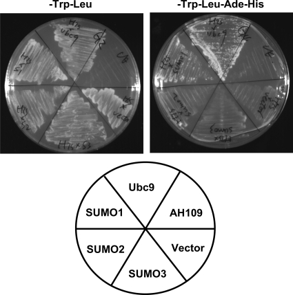
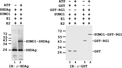
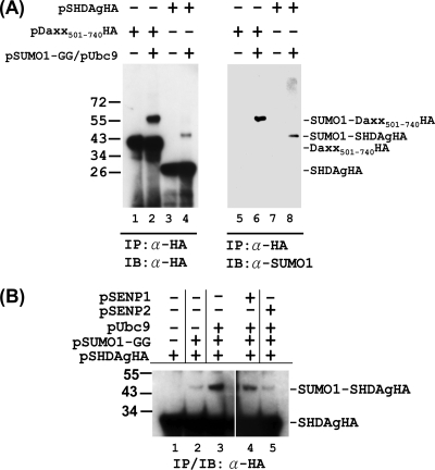
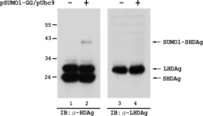
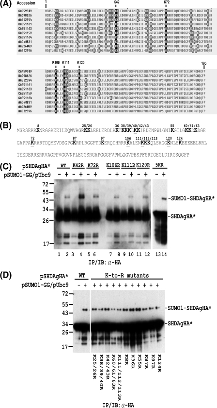
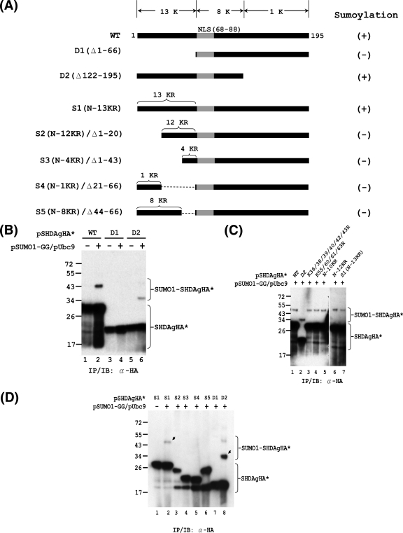
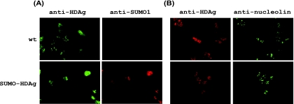
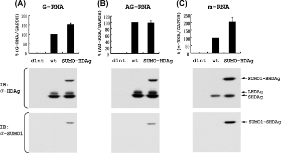
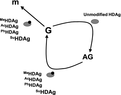
Hepatitis delta antigen (HDAg) is a nuclear protein that is intimately involved in hepatitis delta virus (HDV) RNA replication. HDAg consists of two protein species, the small form (S-HDAg) and the large form (L-HDAg). Previous studies have shown that posttranslational modifications of S-HDAg, such as phosphorylation, acetylation, and methylation, can modulate HDV RNA replication. In this study, we show that S-HDAg is a small ubiquitin-like modifier 1 (SUMO1) target protein. Mapping data showed that multiple lysine residues are SUMO1 acceptors within S-HDAg. Using a genetic fusion strategy, we found that conjugation of SUMO1 to S-HDAg selectively enhanced HDV genomic RNA and mRNA synthesis but not antigenomic RNA synthesis. This result supports our previous proposition that the cellular machinery involved in the synthesis of HDV antigenomic RNA is different from that for genomic RNA synthesis and mRNA transcription, requiring different modified forms of S-HDAg. Sumoylation represents a new type of modification for HDAg.


Figures

FIG. 1.
FIG. 1.
S-HDAg interacts with Ubc9 and SUMO1 in yeast two-hybrid assay. The growth of yeast strain AH109 cotransformed with GAL4-DBD plasmid DNA (pGBKT7−S-HDAg) and different GAL4-AD plasmid DNAs (pACT2-SUMO1, pACT2-SUMO2, pACT2-SUMO3, or pACT2-Ubc9) on SD medium is shown. Cotransformants were isolated on SD medium lacking tryptophan and leucine (−Trp−Leu; left plate) or on SD medium lacking tryptophan, leucine, adenine, and histidine (−Trp−Leu−Ade−His; right plate). The lower panel represents the isolated cotransformants in which the GAL4-AD plasmids were used for cotransformation. Vector, pACT2 plasmid without insert DNA; AH109, DNA-free transformation control.
FIG. 2.
FIG. 2.
Sumoylation of S-HDAg in vitro. The sumoylation system was reconstituted in vitro with recombinant SUMO E1 (Aos1/Uba2), SUMO E2 (Ubc9), SUMO1, and S-HDAg or the control substrate proteins. GST protein and GST-tagged RanGAP1 (GST-RG1) served as negative and positive control substrate proteins, respectively, for this assay (RG1 was the first protein shown to be posttranslationally modified with SUMO [35]). The reactions were carried out in sumoylation buffer with (+) or without (−) ATP. The S-HDAg substrate and the SUMO1-S-HDAg generated in the reaction were analyzed by immunoblotting (IB) with an antibody (α) to HDAg (lanes 1 and 2). The control substrates and their SUMO1-conjugated forms generated in the reaction were analyzed by immunoblotting with an antibody (α) to GST (lanes 3 to 6). The locations of molecular mass markers are indicated on the left, in kDa, and the positions of substrate proteins and their SUMO1-conjugated forms are marked on the right.
FIG. 3.
FIG. 3.
Sumoylation of S-HDAg in vivo. (A) Detection of SUMO1-conjugated S-HDAg in transfected cells. In Huh-7 cells, the expression plasmid for HA-tagged S-HDAg or the Daxx(501-740) truncation mutant was transfected with or without the expression plasmids for SUMO1-GG (the mature form of SUMO1) and Ubc9. The whole-cell extracts were then prepared and subjected to immunoprecipitation (IP) with an antibody to HA tag followed by immunoblotting (IB) with the same antibody or with an antibody to SUMO1, as indicated. Daxx is a well-documented SUMO target protein (30). Transfection of the expression plasmid for Daxx501-740HA served as a positive control for this assay. (B) Overexpression of SENP2 reduces sumoylation of S-HDAg. The expression plasmid for HA-tagged S-HDAg was transfected into Huh-7 cells with or without the expression plasmids for SUMO1-GG, Ubc9, SENP1, and SENP2, as indicated. The whole-cell extracts were then prepared and subjected to IP/IB with an antibody to the HA tag.
FIG. 4.
FIG. 4.
S-HDAg produced from HDV RNA-replicating cells is a SUMO1 target protein. In Huh-7 cells, HDV genomic RNA was transfected together with the S-HDAg-encoding mRNA to establish HDV RNA replication. Four days after RNA transfection, the cells were then transfected again, with or without the SUMO1-GG and Ubc9 expression plasmids. Two days later, the whole-cell extracts were prepared and then subjected to immunoblotting with a mouse monoclonal antibody against HDAg (lanes 1 and 2) or with a rabbit polyclonal antibody specific for L-HDAg (LP3) (49) (lanes 3 and 4).
FIG. 5.
FIG. 5.
Mapping of lysine residues in S-HDAg that are conjugated by SUMO1. (A) Alignment of S-HDAg amino acid sequences from different HDV isolates. The nonconserved and conserved lysine (K) residues are shaded in gray and black, respectively. (B) Amino acid sequence of the Italian HDV isolate used in the in vivo sumoylation assay. The lysine (K) residues are shown in bold. The underlined K residues denote the lysine residues which are conserved among different HDV isolates. (C) The SUMO acceptor lysines within S-HDAg are not limited to the five conserved lysines. In Huh-7 cells, expression plasmids encoding wild-type S-HDAgHA or its mutants containing K-to-R substitutions at the conserved lysine(s) were transfected with or without the SUMO1-GG and Ubc9 expression plasmids. The whole-cell extracts were then prepared and subjected to IP/IB with an antibody to the HA tag. (D) Multiple lysine residues are SUMO1 acceptors within S-HDAg. Expression plasmids encoding wild-type S-HDAgHA or its mutants containing K-to-R substitution(s) were used for in vivo sumoylation assay as in panel C. The positions of the S-HDAgHA species (denoted as SHDAgHA*) and the SUMO1-conjugated form (denoted as SUMO1-SHDAgHA*) are marked on the right. Expression plasmids encoding wild-type S-HDAgHA or its mutants are indicated at the top or bottom.
FIG. 6.
FIG. 6.
Mapping of functional domain required for SUMO modification of S-HDAg. In vivo sumoylation assays were performed with S-HDAg and its mutants. (A) Schematic representation of S-HDAg and its deletion and K-to-R substitution derivatives. The total numbers of lysine (K) residues within the various regions of S-HDAg are indicated at the top. The nuclear localization signal (NLS; amino acids 66 to 88) is shown in gray. The numbers of K-to-R substitutions within the N-terminal region of S1 and its derivatives are indicated on top of these constructs. The ability of the S-HDAg derivatives to undergo sumoylation is summarized on the right. (B) The N-terminally truncated form of S-HDAg loses its ability to be sumoylated. Expression plasmids encoding the wild-type S-HDAgHA (WT) or its deletion mutants, D1 and D2, were used for in vivo sumoylation and subsequent IP/IB assay with anti-HA antibody. (C) The N-terminal 13 lysines are not the exclusive SUMO acceptor lysines of S-HDAg. Expression plasmids encoding wild-type S-HDAgHA and its derivatives (as indicated) were used for in vivo sumoylation and subsequent IP/IB assay with anti-HA antibody. (D) The N-terminal 66 aa of S-HDAg are required for its SUMO modification. The indicated expression plasmids were used for in vivo sumoylation assay as mentioned above. The arrows indicate SUMO1-modified S-HDAgHA* species.
FIG. 7.
FIG. 7.
The SUMO1-S-HDAg fusion protein displays a subcellular localization pattern similar to that of wild-type S-HDAg. The mRNA encoding wild-type S-HDAg (wt) or mutant SUMO1-S-HDAg (SUMO-HDAg) was transfected into Huh-7 cells. At day 1 posttransfection, cells were fixed and prepared for immunofluorescence microscopic examination (as described in Materials and Methods). (A) A mouse anti-HDAg monoclonal antibody or a rabbit anti-SUMO1 polyclonal antibody was used as the first antibody. (B) A rabbit anti-HDAg polyclonal antibody or a mouse anti-nucleolin monoclonal antibody was used as the first antibody.
FIG. 8.
FIG. 8.
Effects of SUMO1 conjugation of S-HDAg on HDV G-RNA, AG-RNA, and mRNA synthesis. HDV AG-RNA (A) or HDV G-RNA (B and C) was cotransfected with an mRNA encoding wild-type S-HDAg (wt), SUMO1-S-HDAg (SUMO-HDAg), or defective S-HDAg (d1nt) into cells to establish HDV RNA replication. At day 4 (A and B) or day 1 (C) posttransfection, total RNAs were extracted, and HDV G-RNA (A), AG-RNA (B), and mRNA (C) were detected by qRT-PCR, using the G-S, AG-S, and mI-S protocols, respectively (48) (upper panels). The data were normalized relative to the values obtained for the wild-type S-HDAg-encoding mRNA cotransfection. Error bars represent standard deviations from the means for three independent experiments. To demonstrate the expression of the expected forms of HDAg (as indicated by arrows), the whole-cell extracts from one of the qRT-PCR assays were prepared and subjected to immunoblotting with a mouse monoclonal antibody to HDAg or SUMO1, as indicated (lower panels).
FIG. 9.
FIG. 9.
Model of HDAg requirement for synthesis of the three HDV RNA species. MeHDAg, AcHDAg, PhHDAg, and SuHDAg represent methylated, acetylated, phosphorylated, and sumoylated HDAg, respectively. m, HDAg-encoding mRNA; G, HDV genomic RNA; AG, HDV antigenomic RNA.

References

    1. Boddy, M. N., K. Howe, L. D. Etkin, E. Solomon, and P. S. Freemont. 1996. PIC 1, a novel ubiquitin-like protein which interacts with the PML component of a multiprotein complex that is disrupted in acute promyelocytic leukaemia. Oncogene 13:971-982. - PubMed
    1. Boggio, R., and S. Chiocca. 2006. Viruses and sumoylation: recent highlights. Curr. Opin. Microbiol. 9:430-436. - PMC - PubMed
    1. Bossis, G., C. E. Malnou, R. Farras, E. Andermarcher, R. Hipskind, M. Rodriguez, D. Schmidt, S. Muller, I. Jariel-Encontre, and M. Piechaczyk. 2005. Down-regulation of c-Fos/c-Jun AP-1 dimer activity by sumoylation. Mol. Cell. Biol. 25:6964-6979. - PMC - PubMed
    1. Carter, S., and K. H. Vousden. 2008. p53-Ubl fusions as models of ubiquitination, sumoylation and neddylation of p53. Cell Cycle 7:2519-2528. - PubMed
    1. Chang, F. L., P. J. Chen, S. J. Tu, C. J. Wang, and D. S. Chen. 1991. The large form of hepatitis delta antigen is crucial for assembly of hepatitis delta virus. Proc. Natl. Acad. Sci. USA 88:8490-8494. - PMC - PubMed

LinkOut - more resources