. 2010 Jan 1;21(1):87-94.
doi: 10.1091/mbc.e09-08-0722. Epub 2009 Nov 4.

Age-dependent preferential dense-core vesicle exocytosis in neuroendocrine cells revealed by newly developed monomeric fluorescent timer protein

Affiliations

Age-dependent preferential dense-core vesicle exocytosis in neuroendocrine cells revealed by newly developed monomeric fluorescent timer protein

Takashi Tsuboi et al. Mol Biol Cell. .

Abstract

Although it is evident that only a few secretory vesicles accumulating in neuroendocrine cells are qualified to fuse with the plasma membrane and release their contents to the extracellular space, the molecular mechanisms that regulate their exocytosis are poorly understood. For example, it has been controversial whether secretory vesicles are exocytosed randomly or preferentially according to their age. Using a newly developed protein-based fluorescent timer, monomeric Kusabira Green Orange (mK-GO), which changes color with a predictable time course, here we show that small GTPase Rab27A effectors regulate age-dependent exocytosis of secretory vesicles in PC12 cells. When the vesicles were labeled with mK-GO-tagged neuropeptide Y or tissue-type plasminogen activator, punctate structures with green or red fluorescence were observed. Application of high [K(+)] stimulation induced exocytosis of new (green) fluorescent secretory vesicles but not of old (red) vesicles. Overexpression or depletion of rabphilin and synaptotagmin-like protein4-a (Slp4-a), which regulate exocytosis positively and negatively, respectively, disturbed the age-dependent exocytosis of the secretory vesicles in different manners. Our results suggest that coordinate functions of the two effectors of Rab27A, rabphilin and Slp4-a, are required for regulated secretory pathway.


Figures

Figure 1.
Figure 1.
Light-absorption properties and excitation and emission spectra of mK-GO. (A) Absorption spectra of mK-GO. (B) pH dependence of the absorption peak at 500 nm and 548 nm. (C) Normalized excitation (broken line) and emission (solid line) spectra of mK-GO. (D) In vitro maturation kinetics: fluorescence emission ratio between 560 nm and 509 nm of freshly synthesized mK-GO were recorded during maturation of the proteins at 37°C.
Figure 2.
Figure 2.
Localization of timer- and mK-GO–tagged dense-core vesicle markers within PC12 cells. TIRF image of PC12 cells showing the distribution of NPY-timer (A), NPY-mK-GO (B), and tPA-mK-GO (C) in 48 h after transfection. Note that the NPY-timer was present throughout the cell cytosol, with the majority located in irregular vesicular structures. (D) Stacked bar chart represents the percent of the number of plasma membrane-docked green, yellow, and red vesicles in NPY- and tPA-mK-GO–expressing cells (n = 28 cells in each). Bar, 10 μm.
Figure 3.
Figure 3.
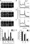
Exocytotic dynamics of dense-core vesicle cargoes NPY and tPA. Five sequential dual color TIRF images show the behavior of single green colored- (A), yellow colored- (C), and red-colored NPY-mK-GO (E) vesicles after applying 70 mM KCl to the cell. The cells were used for experiments in 48 h after transfection. The vesicle position before exocytosis is outlined by a circle. Bars, 1 μm. Fluorescence intensity trances are shown of vesicles containing both green colored- (B), yellow colored- (D), and red colored-NPY-mK-GO vesicles (F). (G) A histogram represents the mean number of plasma membrane-docked NPY-mK-GO vesicles (left; n = 31 cells) or tPA-mK-GO (right; n = 28 cells) vesicles per cell before (closed bars) and after (open bars) high-KCl stimulation. (H) Percentage of the number of NPY- or tPA-mKO release events during 5-min stimulation to the number of plasma membrane-docked vesicle before stimulation. Note that red colored-NPY- or tPA-mK-GO vesicles showed only few exocytotic events. The results are means ± SE.
Figure 4.
Figure 4.
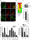
Effect of overexpression or depletion of rabphilin on the dense-core vesicle exocytosis in PC12 cells. (A) Typical TIRF images of plasma membrane-docked green colored- and red colored-NPY-mK-GO vesicles before high-KCl stimulation of scrambled siRNA-transfected control (control), rabphilin-overexpressing (rabphilin), and rabphilin-siRNA-transfected cells (rabphilin-siRNA). The cells were used for experiments in 48 h after transfection. Bar, 10 μm. (B) Stacked bar chart represents the percentage of the number of plasma membrane-docked green, yellow, and red vesicles in control, rabphilin-, and rabphilin-siRNA–transfected cells (n = 35 cells in each). (C) Effect of rabphilin siRNA on expression of rabphilin in PC12 cells. PC12 cells were transfected with rabphilin siRNA (right) or a scrambled-siRNA control (left). Cell lysates were prepared and subjected to 12.5% SDS-PAGE followed by immunoblotting with anti-rabphilin and anti-actin antibodies. The positions of the molecular mass markers (×10−3) are shown on the left. The results shown are representative of three independent experiments with similar results. (D) The density of plasma membrane-docked vesicles was determined by counting the vesicles in each image (n = 31 cells in each). (E). Histograms represent the mean number of plasma membrane-docked NPY-mK-GO vesicles in control (left), rabphilin overexpressing (center), or rabphilin siRNA-transfected cells (right) before (closed bars) and after (open bars) high-KCl stimulation. (F) The effect of expression of rabphilin, or rabphilin siRNA on the percentage of released vesicles. Note that overexpression of rabphilin induced exocytotic events from all colors of NPY-mK-GO vesicles. *p < 0.05 and ***p < 0.001, respectively, in comparison with the control. The results are means ± SE.
Figure 5.
Figure 5.
Effect of overexpression of Slp4-a or Slp4-a-siRNA expression on the dense-core vesicle exocytosis in PC12 cells. (A) Typical TIRF images of plasma membrane-docked green colored- and red colored-NPY-mK-GO vesicles before high-KCl stimulation of scrambled siRNA-transfected control (control), Slp4-a–transfected (Slp4-a), and Slp4-a-siRNA–transfected cells (Slp4- a-siRNA). The cells were used for experiments in 48 h after transfection. Bar, 10 μm. (B) Stacked bar chart represents the percentage of the number of plasma membrane-docked green, yellow, and red vesicles in control, Slp4-a, and Slp4-a-siRNA–transfected cells (n = 25 cells in each). (C) Effect of Slp4-a siRNA on expression of Slp4-a in PC12 cells. PC12 cells were transfected with Slp4-a-siRNA (right) or a scrambled-siRNA control (left). Cell lysates were prepared and subjected to 12.5% SDS-PAGE followed by immunoblotting with anti-Slp4-a and anti-actin antibodies. The positions of the molecular mass markers (×10−3) are shown on the left. The results shown are representative of three independent experiments with similar results. (D) The density of plasma membrane-docked vesicles was determined by counting the vesicles in each image (n = 25 cells in each). (E) Histograms represent the mean number of plasma membrane-docked NPY-mK-GO vesicles in control (left), Slp4-a overexpression (center), or Slp4-a-siRNA–transfected cells (right) before (closed bars) and after (open bars) high-KCl stimulation. (F) The effect of expression of Slp4-a or Slp4-a siRNA on the percentage of released vesicles. Note that Slp4-a siRNA increased the number of plasma membrane-docked green-colored NPY-mK-GO vesicles and facilitated the yellow- and red-colored NPY-mK-GO release events. *p < 0.05, **p < 0.01, and ***p < 0.001, respectively, in comparison with the control. The results are means ± SE.
Figure 6.
Figure 6.
Proposed model for Slp4-a/rabphilin–dependent preferential dense-core vesicle exocytosis in neuroendocrine cells. Rabphilin and Slp4-a promote docking of dense-core vesicles to the plasma membrane through interaction with SNAP-25 (Tsuboi and Fukuda, 2005; Tsuboi et al., 2007) and Munc18-1/syntaxin-1a complex (Tsuboi and Fukuda, 2006b), respectively. Dense-core vesicles docked to the plasma membrane by the rabphilin/SNAP-25 complex (blue bars) undergo preferential exocytosis (presumably corresponds to the readily releasable pool), whereas dense-core vesicles docked to the plasma membrane by the Slp4-a/Munc18-1/sytnaxin-1a complex (red bars) do not undergo exocytosis (corresponds to the reserve pool). The molecular switch between readily releasable pool and reserve pool is currently unknown. Green-, yellow-, and red-colored vesicles correspond to newly synthesized vesicles, middle-aged vesicles, and old vesicles, respectively.

References

    1. Abderrahmani A., Cheviet S., Ferdaoussi M., Coppola T., Waeber G., Regazzi R. ICER induced by hyperglycemia represses the expression of genes essential for insulin exocytosis. EMBO J. 2006;25:977–986. - PMC - PubMed
    1. Axelrod D. Cell-substrate contacts illuminated by total internal reflection fluorescence. J. Cell Biol. 1981;89:141–145. - PMC - PubMed
    1. Baldini G., Martelli A. M., Tabellini G., Horn C., Machaca K., Narducci P., Baldini G. Rabphilin localizes with the cell actin cytoskeleton and stimulates association of granules with F-actin cross-linked by α-actinin. J. Biol. Chem. 2005;280:34974–34984. - PubMed
    1. Cheviet S., Waselle L., Regazzi R. Noc-king out exocrine and endocrine secretion. Trends Cell Biol. 2004;14:525–528. - PubMed
    1. Dannies P. S. Mechanisms for storage of prolactin and growth hormone in secretory granules. Mol. Genet. Metab. 2002;76:6–13. - PubMed

Publication types