. 2010 Jan;48(1):220-8.
doi: 10.1128/JCM.01556-09. Epub 2009 Nov 4.

Neosartorya udagawae (Aspergillus udagawae), an emerging agent of aspergillosis: how different is it from Aspergillus fumigatus?

Affiliations

Neosartorya udagawae (Aspergillus udagawae), an emerging agent of aspergillosis: how different is it from Aspergillus fumigatus?

J A Sugui et al. J Clin Microbiol. 2010 Jan.

Abstract

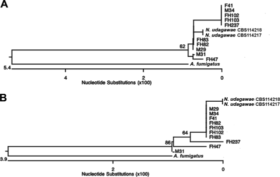
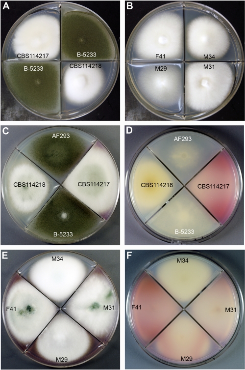
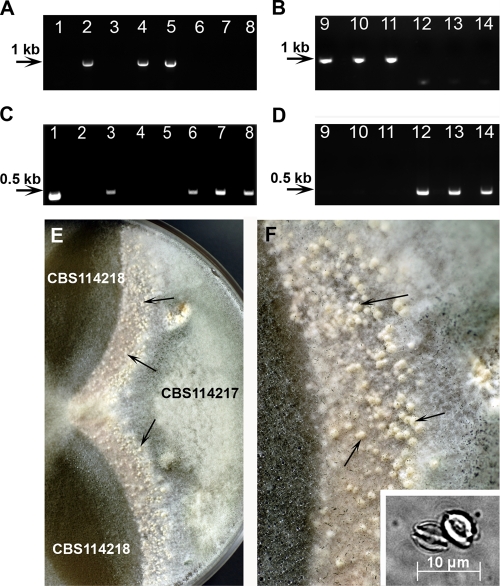
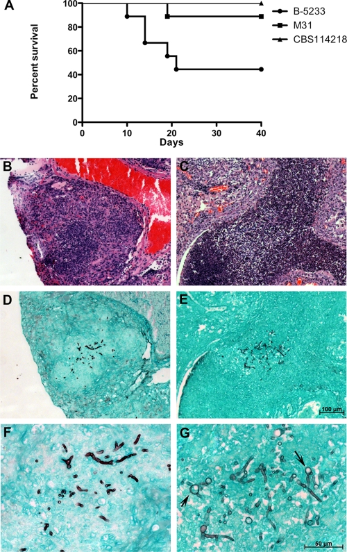
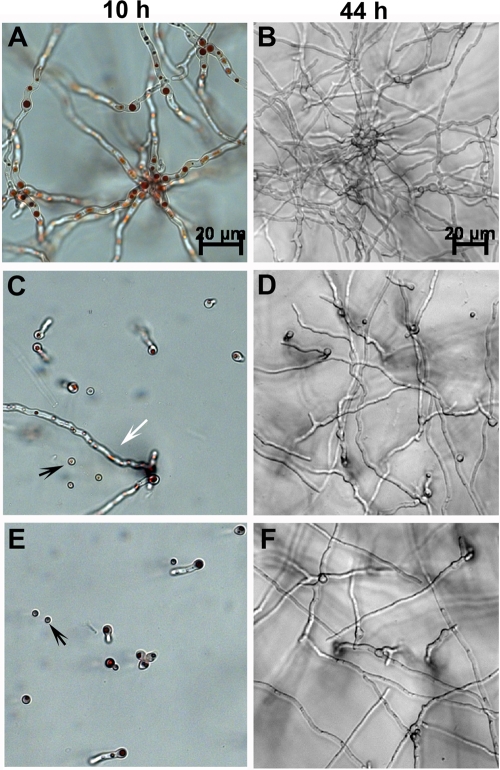
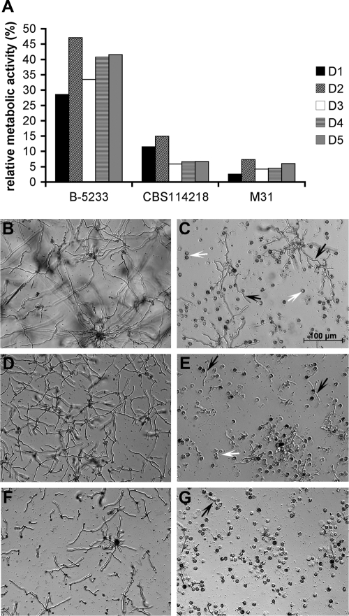
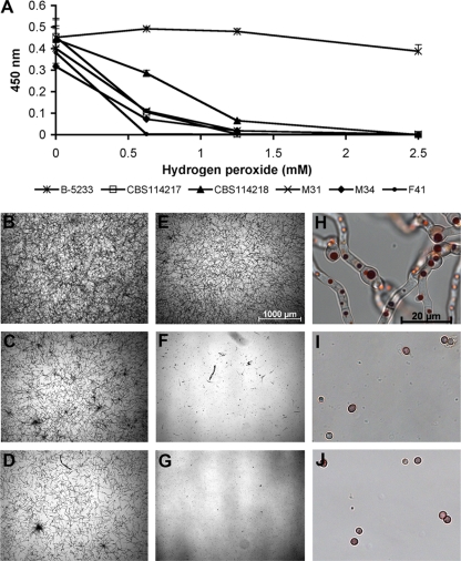
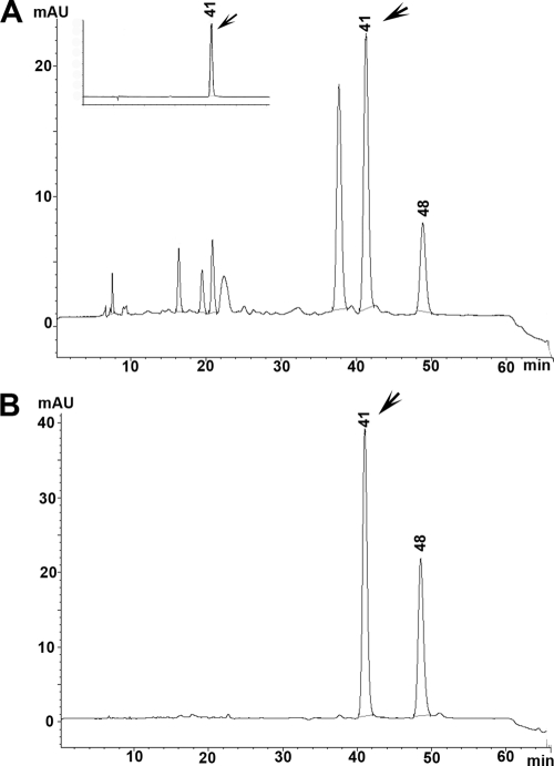
A recent report on several cases of invasive aspergillosis caused by Neosartorya udagawae suggested distinctive patterns of disease progression between N. udagawae and Aspergillus fumigatus. This prompted us to characterize N. udagawae in comparison to A. fumigatus. Our findings showed that both species exist in two mating types at similar ratios and produce gliotoxin. However, the thermotolerance of the two species differs: while A. fumigatus is able to grow at 55 degrees C but not at 10 degrees C, N. udagawae is able to grow at 10 degrees C but fails to grow at >42 degrees C. Furthermore, compared to A. fumigatus, the conidia of N. udagawae require longer incubation periods to germinate at 37 degrees C and are more susceptible to neutrophil attack as well as hydrogen peroxide; N. udagawae is also less virulent in gp91(phox-/-) mice. These findings suggest that growth and susceptibility to the host response might account for the reduced virulence of N. udagawae and the subtle distinction in the progression of the disease caused by the two species.


Figures

FIG. 1.
FIG. 1.
Phylogenetic analysis. Neighbor-joining trees based on partial benA (A) and rodA (B) sequences of clinical and type strains of N. udagawae as well as A. fumigatus are shown. The numbers at the nodes represent bootstrap values (of 1,000 bootstrap replications)of >50%.
FIG. 2.
FIG. 2.
(A and B) Growth characteristics of N. udagawae and A. fumigatus on Czapek-Dox agar after 5 days at 37°C. (A) A. fumigatus strain B-5233 and N. udagawae type strains CBS114217 and CBS114218; (B) N. udagawae clinical strains M29, M31, M34, and F41. (C to F) Growth characteristics of A. fumigatus and N. udagawae on Aspergillus minimal medium after 7 days at 37°C. (C and E) Colony surface; (D and F) reverse surface. A purplish pink compound is evident in the reverse of the colonies of all N. udagawae strains except CBS114218 and M34.
FIG. 3.
FIG. 3.
Genotyping of MAT idiomorphs and mating assay. The mating type was designated on the basis of the presence of the alpha box (MAT1-1) or the HMG domain (MAT1-2) (A to D). (A and B) Amplicons from the HMG domain; (C and D) amplicons from the alpha box. Lanes 1 and 2, A. fumigatus strains B-5233 (MAT1-1) and AF293 (MAT1-2), respectively; lanes 3 and 4, N. udagawae type strains CBS14217 and CBS114218, respectively; lanes 5 to 14, N. udagawae clinical isolates M29 (lane 5), M31 (lane 6), M34 (lane 7), F41 (lane 8), FH47 (lane 9), FH82 (lane 10), FH83 (lane 11), FH102 (lane 12), FH103 (lane 13), and FH237 (lane 14). In the mating assay, strain CBS114217 was crossed with strain CBS114218 on an oatmeal agar plate, and the plate was incubated at 30°C for 18 days (E and F). Arrows point to the cleistothecia produced along the junction between the two strains. Panel F shows a higher magnification of panel E. The inset in panel E shows ascospores released from cleistothecia.
FIG. 4.
FIG. 4.
Virulence assay in a murine model. gp91phox/ mice with CGD were inoculated with A. fumigatus strain B-5233 or the N. udagawae strains CBS114218 and M31. (A) Survival curve; P ≤ 0.01 for CBS114218 compared to B-5233, and P ≤ 0.04 for M31 compared to B-5233 (log-rank test). (B to G). Histopathology of lung tissue from mice that succumbed to infection caused by strains B-5233 (B, D, and F) and M31 (C, E, and G). (B and C) Sections stained with H&E; (D to G) sections stained with GMS. Panels F and G are higher magnifications of panels D and E, respectively. The magnification in panels B to D is the same as that in panel E. The magnification in panel F is indicated in panel G. Arrows indicate swollen round structures formed in the hyphae of N. udagawae strain M31.
FIG. 5.
FIG. 5.
Growth of N. udagawae and A. fumigatus in RPMI 1640 medium. Resting conidia were inoculated in chambered slides containing RPMI 1640 and 25 mM HEPES, and the chambered slides were incubated at 37°C for 10 and 44 h. (A and B) A. fumigatus strain B-5233; (C and D) N. udagawae type strain CBS114218; (E and F) N. udagawae clinical strain M31. The magnification in panels C and E is the same as that in panel A. The magnification in panels D and F is the same as that in panel B. Black arrows, ungerminated conidia; white arrow, occasional conidia that germinated and formed long hyphal filament.
FIG. 6.
FIG. 6.
Effect of human neutrophils on conidia of N. udagawae and A. fumigatus. (A) Metabolic activity of conidia after challenge with neutrophils relative to that of control conidia without neutrophils. Each bar represents a biological replicate carried out with neutrophils from a single donor. Neutrophils from five different donors (donors D1 to D5) were tested. The metabolic activity of the control conidia without neutrophils was considered to be 100%. (B to G) Bright-field microscopy images showing the growth of A. fumigatus B-5233 (B and C) and N. udagawae CBS114218 (D and E) and M31 (F and G). (B, D, and F) Growth in the absence of neutrophils; (C, E, and G) growth in the presence of neutrophils. White arrows, neutrophils; black arrows, hyphal filaments. The magnification indicated in panel C applies to all other panels.
FIG. 7.
FIG. 7.
Effect of hydrogen peroxide on N. udagawae and A. fumigatus. (A) Metabolic activity assayed by the XTT assay and measured at OD450. The conidia of A. fumigatus strain B-5233 and N. udagawae strains CBS114217, CBS114218, M31, M34, and F41 were incubated in the presence of hydrogen peroxide at concentrations of 0.625, 1.25, and 2.5 mM. (B to J) Bright-field microscopy images of B-5233 (B, E, and H), CBS114218 (C, F, and I), and M31 (D, G, and J). (B to D) Conidia incubated without hydrogen peroxide; (E to J) conidia incubated with 2.5 mM hydrogen peroxide. Samples were observed after 20 h incubation. The magnifications in panels H to J are 50 times those in panels E to G. The magnification of panels B to G is indicated in panel E. The magnification bar shown in panel H also applies to panels I and J.
FIG. 8.
FIG. 8.
Analysis of gliotoxin. HPLC analysis of culture supernatants from A. fumigatus strains B-5233 (A) and representative N. udagawae strain M31 (B). The inset in panel A shows the gliotoxin standard. Arrows, the peak that eluted at 41 min, which corresponds to gliotoxin. The identity of the peak eluting at 48 min is unknown. The results of a representative HPLC analysis are shown. mAU, milliabsorbance units.

References

    1. Amitani, R., G. Taylor, E. N. Elezis, C. Llewellyn-Jones, J. Mitchell, F. Kuze, P. J. Cole, and R. Wilson. 1995. Purification and characterization of factors produced by Aspergillus fumigatus which affect human ciliated respiratory epithelium. Infect. Immun. 63:3266-3271. - PMC - PubMed
    1. Balajee, S. A., J. L. Gribskov, E. Hanley, D. Nickle, and K. A. Marr. 2005. Aspergillus lentulus sp. nov., a new sibling species of A. fumigatus. Eukaryot. Cell 4:625-632. - PMC - PubMed
    1. Balajee, S. A., D. Nickle, J. Varga, and K. A. Marr. 2006. Molecular studies reveal frequent misidentification of Aspergillus fumigatus by morphotyping. Eukaryot. Cell 5:1705-1712. - PMC - PubMed
    1. Belkacemi, L., R. C. Barton, V. Hopwood, and E. G. Evans. 1999. Determination of optimum growth conditions for gliotoxin production by Aspergillus fumigatus and development of a novel method for gliotoxin detection. Med. Mycol. 37:227-233. - PubMed
    1. Bonnett, C. R., E. J. Cornish, A. G. Harmsen, and J. B. Burritt. 2006. Early neutrophil recruitment and aggregation in the murine lung inhibit germination of Aspergillus fumigatus conidia. Infect. Immun. 74:6528-6539. - PMC - PubMed

Publication types

MeSH terms

Substances