. 2010 Feb;298(2):C342-54.
doi: 10.1152/ajpcell.00336.2009. Epub 2009 Nov 4.

Protein expression profiling of lens epithelial cells from Prdx6-depleted mice and their vulnerability to UV radiation exposure

Affiliations

Protein expression profiling of lens epithelial cells from Prdx6-depleted mice and their vulnerability to UV radiation exposure

Eri Kubo et al. Am J Physiol Cell Physiol. 2010 Feb.

Abstract

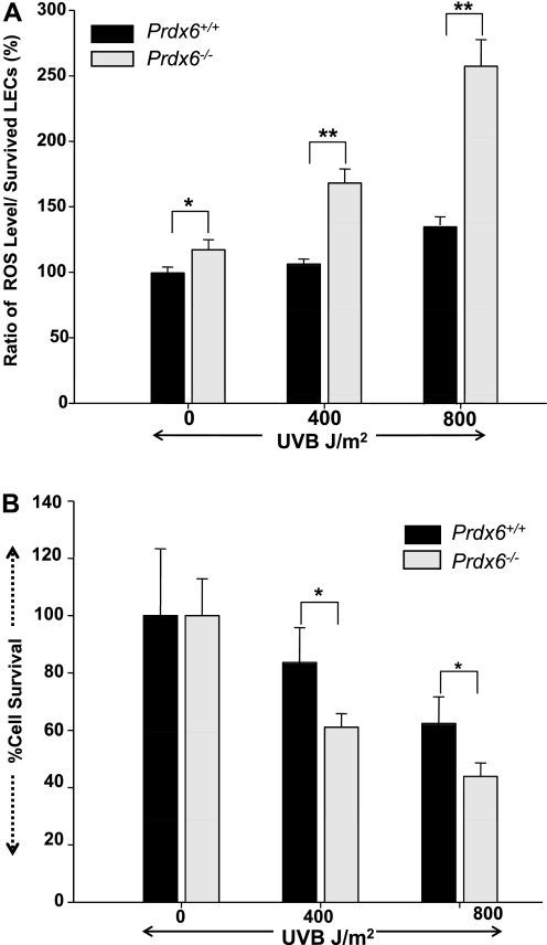
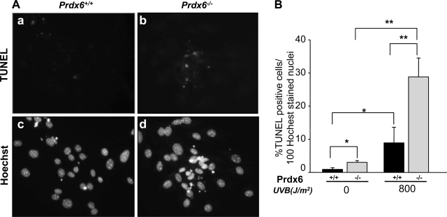
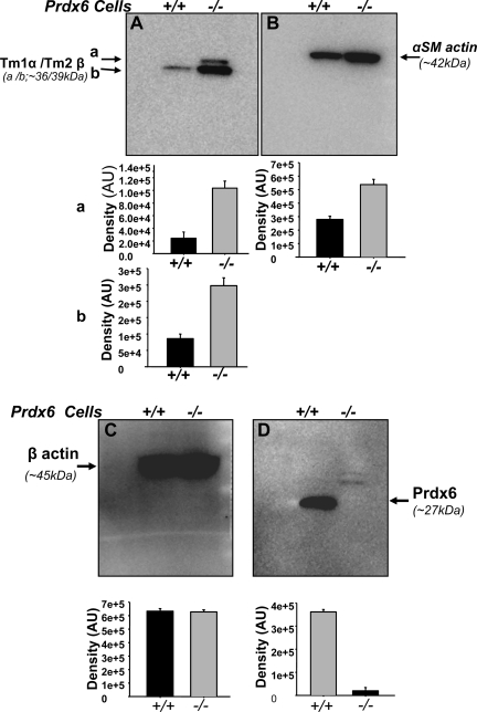
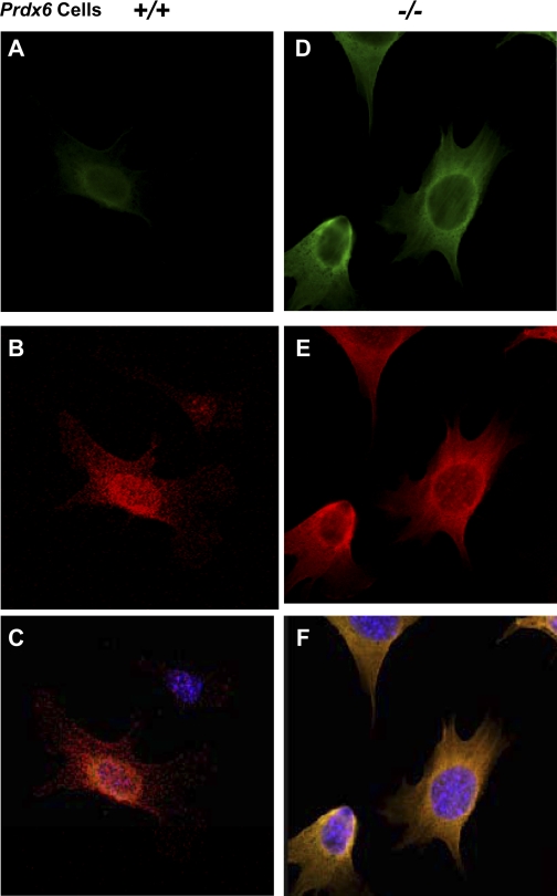
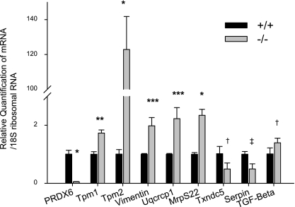
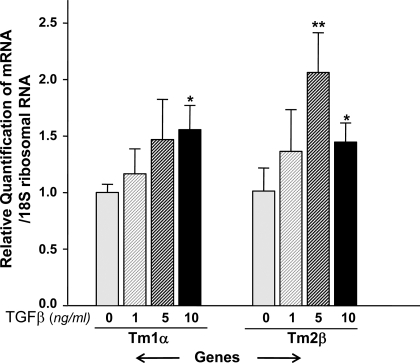
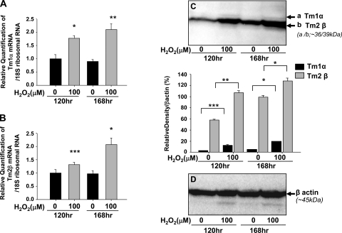
Oxidative stress is one of the causative factors in progression and etiology of age-related cataract. Peroxiredoxin 6 (Prdx6), a savior for cells from internal or external environmental stresses, plays a role in cellular signaling by detoxifying reactive oxygen species (ROS) and thereby controlling gene regulation. Using targeted inactivation of the Prdx6 gene, we show that Prdx6-deficient lens epithelial cells (LECs) are more vulnerable to UV-triggered cell death, a major cause of skin disorders including cataractogenesis, and these cells display abnormal protein profiles. PRDX6-depleted LECs showed phenotypic changes and formed lentoid body, a characteristic of terminal cell differentiation and epithelial-mesenchymal transition. Prdx6(-/-) LECs exposed to UV-B showed higher ROS expression and were prone to apoptosis compared with wild-type LECs, underscoring a protective role for Prdx6. Comparative proteomic analysis using fluorescence-based difference gel electrophoresis along with mass spectrometry and database searching revealed a total of 13 proteins that were differentially expressed in Prdx6(-/-) cells. Six proteins were upregulated, whereas expression of seven proteins was decreased compared with Prdx6(+/+) LECs. Among the cytoskeleton-associated proteins that were highly expressed in Prdx6-deficient LECs was tropomyosin (Tm)2beta. Protein blot and real-time PCR validated dramatic increase of Tm2beta and Tm1alpha expression in these cells. Importantly, Prdx6(+/+) LECs showed a similar pattern of Tm2beta protein expression after transforming growth factor (TGF)-beta or H(2)O(2) treatment. An extrinsic supply of PRDX6 could restore Tm2beta expression, demonstrating that PRDX6 may attenuate adverse signaling in cells and thereby maintain cellular homeostasis. Exploring redox-proteomics (Prdx6(-/-)) and characterization and identification of abnormally expressed proteins and their attenuation by PRDX6 delivery should provide a basis for development of novel therapeutic interventions to postpone ROS-mediated abnormal signaling deleterious to cells or tissues.


Figures

Fig. 1.
Fig. 1.
Photomicrograph of Prdx6−/− lens epithelial cells (LECs) cultured in vitro showing phenotypic changes. Prdx6+/+ (A) and Prdx6−/− (B) LECs were cultured in complete Dulbecco's modified Eagle's medium DMEM containing 10% fetal bovine serum and washed, and the medium was changed to 0.1% bovine serum albumin (BSA) in DMEM. Cells were photomicrographed after 48 h of serum depletion. Under serum-depleted conditions, Prdx6−/− cells became elongated and fiberlike, formed cellular aggregates, packed irregularly, and finally aggregated into lentoid formations and underwent spontaneous apoptosis.
Fig. 2.
Fig. 2.
Prdx6−/− cells showed increased reactive oxygen species (ROS) expression after exposure to UV-B radiation in vitro. Prdx6+/+ and Prdx6−/− cells were cultured for 24 h as described in materials and methods. After culturing, the cells were subjected to UV-B radiation at 0, 400, and 800 J/m2. The medium was replaced 24 h later with Hanks’ solution containing 5–10 μM H2-dichlorofluorescin diacetate (DCFH-DA). Fluorescence intensity (ROS expression) was measured. Histogram in A represents the results, showing elevated expression of intracellular ROS in Prdx6−/− cells compared with Prdx6+/+ cells. *P < 0.006, **P < 0.00001. B: cell survival assay (MTS assay) demonstrating that Prdx6−/− LECs cells are highly vulnerable to UV-B-radiation-induced insults. Cells were cultured as stated above and exposed to UV-B radiation, and an MTS assay was conducted as described in text 24 h later. A significant decrease in cell survival of Prdx6−/− LECs was observed compared with Prdx6+/+ LECs (*P < 0.00015), suggesting that PRDX6 is essential to protect cells from UV-B-radiation-induced damage. Results are means ± SD of 3 independent experiments, and A and B are representative of the experiments.
Fig. 3.
Fig. 3.
Prdx6−/−-deficient cells, which are highly vulnerable to UV-B radiation exposure, undergo apoptosis. Cells were cultured in DMEM + 10% fetal bovine serum (FBS) in 60-mm petri dishes. The day after culturing, cells were exposed to UV-B radiation (800 J/m2). After a 24-h recovery period, cells were stained with either TdT-mediated dUTP-biotin nick end-labeling (TUNEL; A, top) or Hoechst (A, bottom) and photomicrographed. A significant number of apoptotic cell deaths were seen in Prdx6−/− cells (A, b and d). B: % of apoptotic cells/100 Hoechst-stained nuclei. *P < 0.04, **P < 0.001.
Fig. 4.
Fig. 4.
Representative difference gel electrophoresis (DIGE) images of protein extracts of Prdx6−/− and Prdx6+/+ LECs. Labeled samples (Cy3 for Prdx6−/− LECs in green and Cy5 for Prdx6+/+ LECs in red) were subjected to 2-dimensional (2D) gel electrophoresis. Fluorescence was scanned, and the derived images were superimposed with pseudocolors in the dyes. Proteins that are up- or downregulated in Prdx6−/− cells compared with Prdx+/+ LECs are represented by red (up) and green (down) spots, whereas proteins that are equally abundant in both samples appear yellow. The full range of the horizontal axis is from 4 (left) to 7 (right) pH units, and the full range of the vertical axis is from ∼10 (bottom) to ∼100 (top) kDa. A: superimposed images from Cy3- and Cy5-labeled samples in the large gel (13 × 13 cm). Arrows and spot numbers (see Table 1) indicate significant upregulated (red) or downregulated (green) proteins present in Prdx6−/− LECs vs. Prdx6+/+ LECs. B: enlarged area from a small (7 × 7 cm) 2D-SDS gel showing Prdx6−/− (left) and Prdx6+/+ (right) LECs. Protein spots that were decreased or increased >1.5-fold were selected for matrix-assisted laser desorption/ionization time of flight (MALDI-TOF) identification. Mr, molecular weight ratio.
Fig. 5.
Fig. 5.
Western blot analysis of protein extracts from Prdx6−/− and Prdx6+/+ LECs showing upregulation of Tm1α, Tm2β, and α-SMA proteins in Prdx6−/− LECs. The mouse monoclonal Tm Ab (TM311) detects the amino terminal exon 1a of the Tm1α and Tm2β genes and is therefore able to detect recombinant Tm1, -2, -3, and Br-1 (27, 77). Although Tm1 and the β-isoform (36 kDa) from Tm2β gene (A, a) protein were not detected in Prdx6+/+ LECs, they were detected in Prdx6−/− LECs. Tm1 or β (A, a = 36 kDa), and Tm2 or α (A, b = 39 kDa) isoforms from Tm2β and Tm1α genes, respectively, and α-SMA proteins (B) were increased in Prdx6−/− LECs. No change was observed in β-actin expression (C, compare black vs. gray bars), confirming equal protein loading and suggesting that expression modulation for other proteins is specific. D confirms that LECs Prdx6−/− LECs do not express PRDX6 protein. Histograms represent densities of protein bands. Results are derived from 3 different cell preparations. AU, arbitrary units.
Fig. 6.
Fig. 6.
Confocal laser scanning images of Prdx6+/+ and Prdx6−/− LECs showing Tm1α/2β immunolocalization and F-actin. F-actin was labeled with Texas Red in both Prdx6+/+ (B) and Prdx6−/− (E) LECs. Tm1α/2β protein was strongly stained in Prdx6−/− LECs (D) compared with Prdx6+/+ LECs (A) after administration of anti-Tm1α/2β Ab and use of an Alexa Fluor 488 Signal-Amplification Kit. Tm1α/2β proteins can be seen to be localized to stress fiber formed in Prdx6−/− LECs (B). C and F: merged images of Tm immunostaining and F-actin staining.
Fig. 7.
Fig. 7.
Quantitative real-time PCR analysis of Prdx6−/− and Prdx6+/+ LEC mRNA showing expression of Tm1α, Tm2β, Vimentin, Uqcrcp1, MrpS22, Txndc5, Serpinb6a, and TGF-β1. Total RNA was isolated and transcribed into cDNA. Real-time PCR was conducted with specific primers corresponding to genes (see materials and methods). mRNA expression of each gene was adjusted to the 18S ribosomal RNA. Results showed that Tm1α, Tm2β, Vimentin, Uqcrcp1, MrpS22, and TGF-β1 mRNA were upregulated and Txndc5 and Serpinb6a mRNA were downregulated in Prdx6−/− LEC. *P < 0.000, **P < 0.0008, ***P < 0.005, †P < 0.05, ‡P < 0.001.
Fig. 8.
Fig. 8.
Real-time PCR analysis of Prdx6+/+ LEC mRNA with or without addition of transforming growth factor (TGF)-β1, showing TGF-β1-mediated regulation of Tm1α and Tm2β. Cells were cultured in petri dishes in DMEM containing 10% FBS. After 24 h, cells were washed and treated with varying concentrations of TGF-β1 containing 0.2% BSA for 48 h or more. After incubation, RNA was isolated and real-time PCR was conducted. Treatment of Prdx6+/+ LECs with 5.0 or 10.0 ng/ml of TGF-β1 resulted in induction of Tm1α and Tm2β (*P < 0.05, **P < 0.015), suggesting that upregulation of these genes is associated with activation and expression of TGF-β1 in Prdx6−/− cells, as reported elsewhere (21).
Fig. 9.
Fig. 9.
Real-time PCR analysis of Prdx6−/− LECs mRNA supplied with PRDX6 (TAT-HA-PRDX6) protein revealing attenuation of overactivation of Tm2β genes. Cells were cultured in the presence or absence of 10 μg/ml of recombinant TAT-HA-linked Prdx6 protein, as described previously (41). Addition of TAT-HA-Prdx6 protein significantly suppressed overproduction of Tm2β mRNA in Prdx6−/− LECs. *P < 0.05.
Fig. 10.
Fig. 10.
Real-time PCR and protein blot analysis of Prdx6+/+ LECs mRNA with or without addition of H2O2 showing ROS regulation of Tm1α and -2β. Prdx+/+ LECs were cultured in the presence or absence of 100 μM H2O2 for 120 and 168 h. Quantitative real-time PCR (A, B) and protein blotting (C, D) were performed to analyze the expression level of Tm1α and Tm2β transcripts and proteins. Expression of Tm1α and Tm2β transcripts (A) and proteins (B) were significantly increased in Prdx+/+ LECs treated with H2O2. *P < 0.002, **P < 0.0001, ***P < 0.028.

References

    1. Ames BN, Shigenaga MK, Hagen TM. Oxidants, antioxidants, and the degenerative diseases of aging. Proc Natl Acad Sci USA 90: 7915–7922, 1993 - PMC - PubMed
    1. Andley UP, Clark BA. The effects of near-UV radiation on human lens beta-crystallins: protein structural changes and the production of O2- and H2O2. Photochem Photobiol 50: 97–105, 1989 - PubMed
    1. Annes JP, Munger JS, Rifkin DB. Making sense of latent TGFbeta activation. J Cell Sci 116: 217–224, 2003 - PubMed
    1. Arita T, Lin LR, Reddy VN. Differentiation of human lens epithelial cells in tissue culture. Exp Eye Res 47: 905–910, 1988 - PubMed
    1. Arita T, Lin LR, Susan SR, Reddy VN. Enhancement of differentiation of human lens epithelium in tissue culture by changes in cell-substrate adhesion. Invest Ophthalmol Vis Sci 31: 2395–2404, 1990 - PubMed

Publication types

MeSH terms

LinkOut - more resources